")

If you’re planning cataract surgery, you’ve probably heard a lot about lens implants, anaesthesia choices, and recovery timelines. But one crucial area that often gets overlooked—especially in complex cases—is the condition of the back of the eye, specifically the vitreomacular interface (VMI). Disorders in this region, like epiretinal membranes (ERM) or vitreomacular traction (VMT), can quietly compromise your visual outcome, even if the surgery itself goes smoothly.
In this article, we’re going to unpack how these pre-existing VMI disorders affect your cataract surgery results. We’ll dive into what the latest OCT-based studies show, explain what it means if you’ve been diagnosed with ERM or VMT, and discuss what you can do to optimise your post-op vision.
Understanding the Vitreomacular Interface: What Are We Dealing With?
The vitreomacular interface is where the vitreous body—the jelly-like substance filling most of your eyeball—meets the macula, which is the part of your retina responsible for sharp central vision. This interface is incredibly important because it’s a point of structural tension. Over time, or due to various eye conditions, changes can occur that disrupt this delicate balance.
Two common disorders you might hear about are:
- Epiretinal Membrane (ERM): This is a thin, fibrous layer that forms on the surface of the macula. It can contract over time and cause the retina to wrinkle, leading to distorted or blurry vision.
- Vitreomacular Traction (VMT): Here, the vitreous gel remains partially attached to the macula and exerts traction. This pulling can lead to macular deformation, oedema, or even a macular hole in severe cases.
These conditions can occur silently and often coexist with cataracts in older adults. The problem? They may limit how much your vision improves after cataract surgery—even if everything else goes perfectly.
Why This Matters Before Cataract Surgery
If you’re dealing with a cataract, your surgeon will focus on replacing the clouded lens with a clear intraocular lens (IOL). But if you also have a VMI disorder, your retina may not be in the best shape to make full use of that new clarity. Essentially, it’s like putting a brand-new lens on a slightly warped camera sensor—it helps, but the image still won’t be perfect.
This is where things get clinically interesting. Many patients assume cataract surgery is a standalone procedure, but in cases with coexisting retinal issues, the eye must be considered holistically. An ERM, for example, might not cause major problems before surgery but could become more symptomatic after, when the cataract no longer masks the retinal distortion.
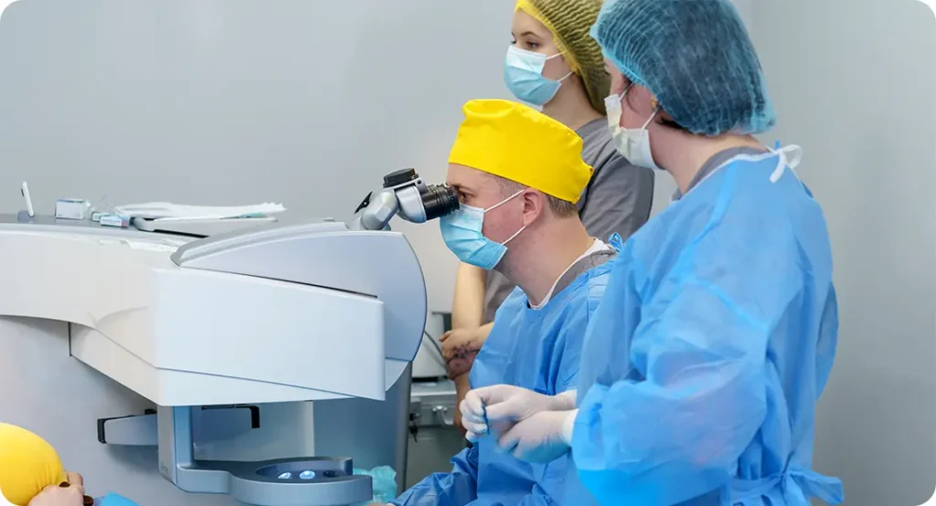
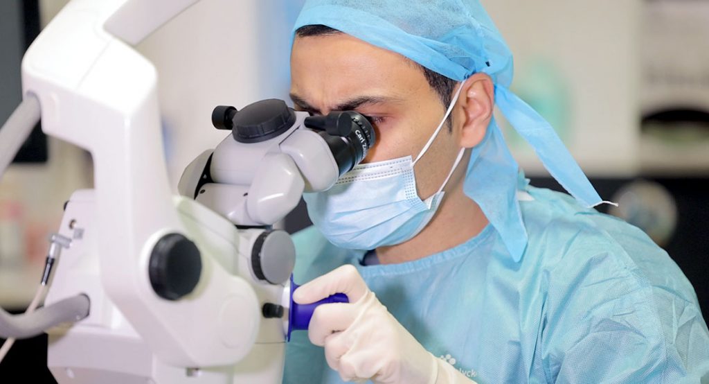
OCT Imaging: The Gold Standard for Preoperative Assessment
Optical Coherence Tomography (OCT) has become a game-changer in cataract surgery planning. It gives us high-resolution, cross-sectional images of the retina—especially the macula—and can reveal early or subtle signs of VMI disorders that are invisible during a routine eye exam.
If you’re scheduled for cataract surgery, your ophthalmologist may suggest an OCT scan to rule out (or confirm) these disorders. It’s a painless and quick test, but its impact is huge. A preoperative OCT can guide the surgical approach, IOL choice, and help predict your post-op visual recovery.
Some surgeons will defer cataract surgery in favour of addressing the VMI issue first—especially in cases of severe VMT or ERM causing macular thickening or cystoid macular oedema (CMO). In other situations, the cataract surgery proceeds first, with careful monitoring post-op.
Epiretinal Membranes and Cataract Surgery: What the Research Shows
A number of studies using OCT data have looked into how ERMs affect cataract outcomes. The general consensus is this: ERMs can reduce the final best-corrected visual acuity (BCVA) after cataract surgery, even when the procedure goes well.
Key Findings from the Literature
- Visual Acuity Gains Are Often Limited: Patients with pre-existing ERMs often report suboptimal improvement in vision, particularly if the ERM is thick or causes significant retinal distortion.
- Visual Distortion Persists: Symptoms like metamorphopsia (straight lines appearing wavy) often don’t resolve post-surgery, because they stem from retinal wrinkling rather than lens opacity.
- Macular Oedema Is More Likely: Postoperative swelling in the retina, or cystoid macular oedema (CMO), is more common in eyes with ERM and can further dampen visual recovery.
One large-scale retrospective study even suggested that delaying ERM peel surgery in patients undergoing cataract removal can lead to poorer long-term results, compared to those who had both issues managed simultaneously or in sequence.
Vitreomacular Traction and Cataract Surgery Outcomes
VMT presents a different challenge. The pulling effect on the macula can distort its architecture, sometimes irreversibly. When left untreated, it may lead to the formation of a full-thickness macular hole—a serious complication.
Key Points from OCT-Based Research
- VMT Can Worsen Post-Surgery: Some cases of VMT resolve spontaneously after cataract surgery, due to vitreous changes triggered by the procedure. However, in others, the traction worsens and leads to visual decline.
- Macular Holes May Develop: Although rare, there are reported cases where cataract surgery appears to accelerate the formation of macular holes in eyes with pre-existing VMT.
- Intraocular Inflammation Can Exacerbate VMT: Cataract surgery induces intraocular inflammation, which may destabilise pre-existing tractional forces at the macula.
If VMT is noted preoperatively, your surgeon may consider combining your cataract operation with a vitrectomy (where the vitreous gel is removed to relieve traction), or stage the surgeries carefully to avoid complications.
How These Disorders Affect IOL Selection
Choosing the right IOL is a personalised decision, but the presence of VMI abnormalities can influence what’s best for you.
- Premium Lenses May Not Be Ideal
Multifocal IOLs or extended depth-of-focus (EDOF) lenses split light to give you both distance and near vision. But they depend heavily on a pristine retinal surface. If your macula is compromised by ERM or VMT, the reduced contrast sensitivity and visual distortion these lenses sometimes cause can be amplified, leaving you less satisfied than expected. - Monofocal IOLs Often Provide More Predictability
For patients with VMI disorders, monofocal lenses focused for distance often deliver the most stable outcomes. They offer excellent clarity, minimal visual aberrations, and are less reliant on a “perfect” macula to function well.
When to Consider Combined or Sequential Surgery
Not every patient with a VMI disorder needs immediate retinal surgery. But in certain cases, managing the retinal condition before or at the same time as cataract surgery can dramatically improve outcomes.
- Combined Surgery
This involves performing cataract extraction and vitrectomy (with ERM peel or VMT release) in a single operation. It’s efficient and avoids multiple rounds of anaesthesia and recovery, but it comes with added complexity and a slightly longer healing period. - Sequential Surgery
In this approach, one issue is tackled first—usually the retinal disorder—followed by cataract surgery a few months later. This staged method is often safer for eyes with fragile maculas or those at risk of macular hole formation.
Patient Counselling: Setting Realistic Expectations
One of the most important aspects of managing cataract patients with VMI disorders is communication. Your surgeon should walk you through:
- The likely visual outcomes given your retinal status
- The risks of leaving the VMI untreated
- Whether you’re a good candidate for a combined approach
- Why certain IOLs might not be appropriate in your case
Visual improvement is still very likely in most cases, but it may not reach the same level as in someone with a healthy retina. The goal is to maximise functional vision while avoiding unnecessary complications.
The Role of the London Cataract Centre in Managing Complex Cases
If you’re considering cataract surgery in London and you know—or suspect—you may have a retinal condition like ERM or VMT, the London Cataract Centre is equipped to help. With access to advanced OCT diagnostics, collaborative care between cataract and retinal surgeons, and a strong emphasis on personalised treatment planning, your surgery can be tailored to the unique anatomy of your eye.
Don’t leave things to chance. Book a consultation that includes a full retinal assessment—because great vision is about more than just a clear lens.
Frequently Asked Questions
- Can I still have cataract surgery if I have an epiretinal membrane (ERM)?
Yes, you can still undergo cataract surgery if you have an ERM, but the presence of the membrane may affect your final visual outcome. While surgery will likely improve your vision by removing the cloudy lens, the retinal distortion caused by the ERM might still result in blurred or distorted vision afterwards. Your surgeon will assess the thickness and location of the membrane using OCT imaging, and may recommend combined or staged surgery in some cases. - Will vitreomacular traction (VMT) go away after cataract surgery?
In some cases, VMT may resolve spontaneously following cataract surgery due to the changes in vitreous dynamics triggered by the operation. However, this is not guaranteed. For others, the traction may worsen, potentially leading to complications like macular holes or increased distortion. Your ophthalmologist will use OCT imaging preoperatively to determine the risk and guide the best surgical strategy. - Should I avoid multifocal lenses if I have a VMI disorder?
Generally, yes. Premium IOLs like multifocal or EDOF lenses work best when the retina is healthy and distortion-free. If you have an ERM or VMT, you may experience suboptimal results with these lenses, including reduced contrast sensitivity and visual disturbances. In such cases, monofocal IOLs are typically a safer and more reliable option, offering clearer and more stable vision. - Can cataract surgery worsen a pre-existing VMI disorder?
It can in some cases. Although cataract surgery itself does not directly damage the retina, it induces intraocular inflammation and changes in the vitreous body. This may destabilise a VMT or make an ERM more symptomatic. Postoperative OCT monitoring is important in patients with pre-existing VMI disorders to catch and manage any changes early. - Is it better to have retinal surgery before or after cataract surgery?
This depends on the severity of the VMI disorder and the degree of visual impairment from the cataract. In cases where the retinal issue is advanced, such as thick ERMs or significant VMT with macular distortion, treating the retina first may provide a better visual foundation for cataract surgery later. However, when the cataract is very dense, it may need to be removed first to allow a clearer view of the retina for any future surgery. - How is a vitreomacular interface disorder diagnosed before cataract surgery?
The gold standard for diagnosing VMI disorders is an OCT (Optical Coherence Tomography) scan. This non-invasive imaging test provides a high-resolution cross-section of your retina, revealing subtle traction or membrane formations that might not be visible during a standard eye exam. Your ophthalmologist will review the OCT images to assess the presence, severity, and clinical impact of any vitreomacular changes before deciding on the best surgical plan. - Can vitreomacular interface disorders cause vision problems even without a cataract?
Yes, they can. Disorders like ERM or VMT can distort central vision, causing symptoms such as blurriness, wavy lines (metamorphopsia), or difficulty reading—even if your lens is still clear. In fact, many people with VMI disorders notice their vision isn’t right, but assume it’s due to a cataract, when the issue is actually retinal. That’s why it’s so important to have the macula checked thoroughly before planning cataract surgery. - Will my recovery be longer if I have a VMI disorder?
Not necessarily, but the visual recovery may be slower or less complete if the retinal surface is compromised. The healing time for the cataract itself is usually unaffected, but your final visual clarity could take longer to stabilise, especially if the VMI disorder progresses or if macular swelling occurs post-op. Regular follow-up and possible use of anti-inflammatory drops or additional treatments will help support your recovery.
Final Thoughts
The interplay between the vitreomacular interface and cataract surgery outcomes is a nuanced one. ERMs and VMTs don’t always prevent a good result—but they do introduce variables that demand careful planning. With proper imaging, open communication, and a surgical plan that respects the underlying retinal condition, you can still achieve excellent functional vision.
If you’re dealing with both a cataract and a retinal condition, don’t assume they must be treated separately or in a rushed timeline. Let your ophthalmologist guide you through a step-by-step approach that prioritises your long-term vision. After all, the real goal isn’t just to see more clearly—but to see with confidence and comfort for years to come.
References
- Govetto, A. et al. (2020) ‘Epiretinal Membranes: New Classification System and Surgical Outcomes’, British Journal of Ophthalmology, 104(6), pp. 839–844.
- Duker, J.S. et al. (2013) ‘The International Vitreomacular Traction Study Group Classification of Vitreomacular Adhesion, Traction, and Macular Hole’, Ophthalmology, 120(12), pp. 2611–2619.
- Johnson, M.W. (2021) ‘Diagnosis and Management of Vitreomacular Interface Disorders’, Retina Today, May 2021, pp. 44–49. Available at: https://retinatoday.com
- Rizzo, S. et al. (2012) ‘Combined Phacoemulsification and Vitrectomy Versus Sequential Surgery in Epiretinal Membrane Patients’, Graefe’s Archive for Clinical and Experimental Ophthalmology, 250(6), pp. 845–852.
- Liu, L. et al. (2022) ‘Impact of Epiretinal Membrane on Visual Outcomes Following Cataract Surgery’, Eye, 36(4), pp. 780–787.