")

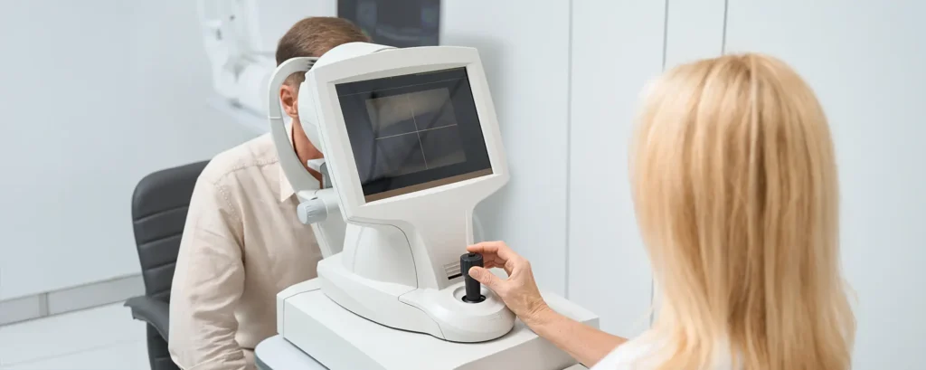
When most people think about cataract surgery, they imagine the surgeon simply removing a cloudy lens and replacing it with a clear implant. What often goes unnoticed is the amount of detailed planning and precision required behind the scenes to make the surgery as safe and predictable as possible. Imaging is at the heart of that process, and in recent years, ultrasound technology has taken on a far more advanced role than the simple scans used decades ago.
Traditionally, ultrasound was associated with older A-scan biometry techniques, where sound waves were bounced off the internal structures of the eye to estimate lens power. Today, however, new high-resolution ultrasound systems are providing incredibly detailed pictures of the eye’s anatomy—sometimes even in situations where optical methods like OCT (optical coherence tomography) and optical biometry fall short. This is especially valuable for patients with dense cataracts, corneal scarring, or other complex conditions where light-based imaging struggles to penetrate.
So in this article, we’re going to take a deep dive into how ultrasound imaging has evolved, why it matters for cataract surgery, and how it works alongside modern diagnostic tools to give surgeons and patients the very best outcomes.
A Brief History of Ultrasound in Ophthalmology
Ultrasound has been used in eye care since the 1950s, originally adapted from medical ultrasound machines used in other areas of the body. The principle is simple: sound waves are transmitted into the eye, and when they bounce back, the echoes are converted into images or measurements. Early versions were quite basic, focusing mainly on axial length measurements (the distance from the cornea to the retina), which are critical for calculating the power of intraocular lenses (IOLs).
For years, A-scan ultrasound was the standard for IOL power calculations. It was effective, but it had its drawbacks: it relied on precise probe placement, could be influenced by operator technique, and wasn’t always as accurate as surgeons would have liked. With the advent of optical biometry in the 1990s and early 2000s, ultrasound fell somewhat into the background, as light-based measurements offered greater precision and reproducibility for routine cases.
But ultrasound never disappeared. Instead, it was being refined in the background. B-scan ultrasound, which produces cross-sectional images of the eye and orbit, became essential for detecting posterior segment issues when the view was obscured by cataracts or vitreous haemorrhage. And now, with high-frequency ultrasound biomicroscopy (UBM) and newer anterior segment ultrasound devices, the technology has staged a remarkable comeback, providing unique insights that other imaging methods can’t always deliver.
Why Ultrasound Still Matters in the Age of OCT and Optical Biometry
It’s tempting to assume that optical technology has replaced ultrasound altogether. After all, devices like the IOLMaster and Lenstar have become the gold standard for lens measurements in routine cataract cases. OCT, meanwhile, has transformed how we look at the retina and macula, providing micrometre-level resolution.
Yet ultrasound continues to hold an important role, precisely because it doesn’t rely on clear optical pathways. In patients with mature or hypermature cataracts, dense corneal opacities, or vitreous haemorrhage, light can’t penetrate well enough to give reliable results. Ultrasound, however, uses sound waves, which pass through opacities that would otherwise block optical systems.
This means ultrasound can provide critical measurements even in the most challenging eyes. It can reveal the condition of the posterior capsule, detect zonular instability, and identify any pathology in the retina or vitreous that might influence the surgical approach. Far from being outdated, it remains a cornerstone in preoperative assessment when optical tools reach their limits.
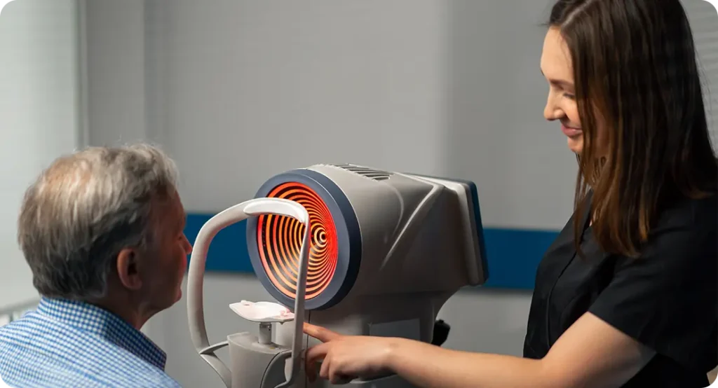
High-Resolution Ultrasound Biomicroscopy (UBM)
One of the biggest advances in ultrasound for cataract surgery is ultrasound biomicroscopy (UBM). Unlike traditional ultrasound, which uses lower frequencies (around 10 MHz), UBM uses very high frequencies (35–100 MHz), allowing for incredibly detailed imaging of the anterior segment of the eye.
With UBM, surgeons can visualise the structures around the lens in remarkable detail: the ciliary body, the iris, the angle, and the zonules that hold the lens in place. This is invaluable in cases where there is suspicion of zonular weakness, trauma, or pseudoexfoliation syndrome. By having a clearer picture of the supporting structures before surgery, the surgeon can plan whether additional devices, such as capsular tension rings, may be required.
UBM also helps in eyes that have had previous surgery, such as trabeculectomy or glaucoma drainage devices, where knowing the position and condition of the tissues is essential for avoiding complications. It has even been used to identify hidden cysts or tumours that could otherwise complicate surgery.
3D and Quantitative Ultrasound Imaging
Another exciting development is the shift from 2D cross-sectional images to 3D reconstructions. Advanced ultrasound systems can now generate volumetric images of the anterior segment, giving surgeons a more complete picture of the lens capsule, cornea, and angle structures. This three-dimensional data makes it easier to appreciate spatial relationships that may influence surgical planning.
Quantitative ultrasound is also on the rise. Instead of just providing images, some systems now measure tissue characteristics like reflectivity and density. This data can be used to evaluate the hardness of the cataract lens, potentially helping the surgeon decide how much energy will be required during phacoemulsification. That means better planning, shorter surgery times, and reduced risk of complications like corneal endothelial cell loss.
Complementing Optical Biometry
Rather than competing with optical biometry, ultrasound often works in partnership with it. In most modern cataract practices, optical biometry is the first choice for IOL calculations. But if the optical device fails to acquire accurate data, ultrasound steps in as a reliable backup.
For example, in a patient with a dense posterior subcapsular cataract, the optical system may struggle to capture the full axial length. An A-scan ultrasound, whether contact or immersion, can fill in that gap. Similarly, if keratometry readings are available from optical devices but axial length is not, combining both sources of data ensures that calculations remain accurate.
By blending optical and ultrasound data, surgeons can cross-verify results and feel more confident in their IOL selection, especially in borderline or complex cases.
Intraoperative Ultrasound Applications
Ultrasound isn’t just for preoperative planning—it’s also finding a role during surgery itself. Intraoperative ultrasound probes, integrated into surgical microscopes or handheld devices, allow real-time visualisation of structures as the procedure unfolds.
This is particularly helpful in cases with poor red reflex or compromised capsule visibility. In such situations, ultrasound can confirm the position of the IOL, guide the removal of residual lens fragments, and help avoid damage to delicate structures. Some experimental systems are even exploring real-time phacoemulsification guidance, where ultrasound maps the lens density dynamically during surgery.
The idea is simple: the more information the surgeon has at their fingertips during surgery, the safer and smoother the operation becomes.
Role in Complex Cataract Cases
Not all cataracts are straightforward. Some patients present with unusual anatomy, previous ocular trauma, high myopia, or conditions like Marfan syndrome that make the lens capsule and zonules more fragile. In these cases, standard imaging may not reveal the full extent of the risks.
Ultrasound biomicroscopy can highlight zonular weakness that might otherwise be missed, while B-scan ultrasound can check the retina for detachment or tumours hidden behind a dense cataract. In highly myopic eyes, ultrasound provides reassurance about the integrity of the posterior segment before surgery.
These insights aren’t just academic—they directly influence surgical choices. For example, detecting zonular laxity ahead of time means the surgeon can be prepared with capsular hooks or tension rings. Spotting a retinal detachment before cataract surgery means the patient can be referred for retinal repair before lens removal, preventing avoidable complications.
Comparing Ultrasound with OCT
It’s worth drawing a direct comparison between ultrasound and OCT, as both technologies are widely used in ophthalmology. OCT uses light waves to generate detailed images, particularly of the retina and macula, and is superb for detecting subtle pathology. Its resolution is far higher than ultrasound, often down to a few microns.
However, OCT is limited by the need for clear optical media. It struggles with dense cataracts or corneal scars. Ultrasound, in contrast, has lower resolution but penetrates even through opaque tissues. This makes it complementary rather than competitive.
In practice, most cataract surgeons now use both: OCT to assess macular health in routine cases, and ultrasound to provide critical backup in cases where the optical view is compromised. By combining both, patients get the best of both worlds.
Training and Accessibility
One of the challenges with ultrasound is that it requires skill to perform and interpret correctly. Optical biometry is relatively straightforward—patients simply sit at a machine and the device captures data automatically. Ultrasound, particularly A-scan immersion or UBM, is more operator-dependent.
That said, training has improved significantly. Ophthalmic technicians and surgeons can now be trained quickly using simulation tools, and new machines are becoming more user-friendly with automated alignment and digital recording. Accessibility is also improving: portable ultrasound devices are making it easier to use this technology even in smaller clinics or mobile surgical settings.
The Future of Ultrasound in Cataract Surgery
Looking ahead, ultrasound is likely to continue evolving in exciting directions. Higher frequency probes may push resolution further, while integration with artificial intelligence could help automate interpretation. Imagine a system that not only provides an image but also flags areas of zonular weakness or predicts lens hardness with a confidence score.
There’s also potential for hybrid systems that combine ultrasound and OCT in one platform, offering the best of both technologies simultaneously. And as cataract surgery itself becomes more advanced—think femtosecond lasers, premium IOLs, and adjustable lenses—the need for equally advanced imaging will only grow.
FAQs
1. What role does ultrasound imaging play in cataract surgery?
Ultrasound imaging plays a vital role in both planning and carrying out cataract surgery. It allows the surgeon to measure the eye’s internal structures, particularly axial length, which is essential for calculating the correct intraocular lens (IOL) power. Beyond measurements, ultrasound can visualise the condition of the lens capsule, the zonules that hold the lens in place, and the retina behind the cataract. These details help identify potential risks before surgery begins. In eyes where optical imaging cannot be performed, ultrasound becomes the main source of accurate anatomical information, making it indispensable in complex or advanced cataract cases.
2. How is ultrasound different from OCT for cataract patients?
Optical coherence tomography (OCT) and ultrasound are often compared because both create cross-sectional images of the eye, but they work in very different ways. OCT uses light waves and provides incredibly high-resolution images—down to microns—making it excellent for examining the retina and macula. However, light cannot penetrate through dense cataracts or corneal scars, meaning OCT fails in many patients who need surgery the most. Ultrasound, on the other hand, relies on sound waves, which travel easily through opaque tissue. Although its resolution is lower than OCT, it has the advantage of working in almost any condition, ensuring that surgeons can still gather critical information even when the optical path is blocked.
3. What exactly is ultrasound biomicroscopy (UBM)?
Ultrasound biomicroscopy (UBM) is a specialised form of ultrasound that uses very high-frequency sound waves, usually between 35 and 100 MHz, to capture highly detailed images of the eye’s anterior segment. With this technique, surgeons can clearly see the iris, ciliary body, angle structures, and the zonules that support the lens. This level of detail is particularly useful in planning cataract surgery for patients with trauma, pseudoexfoliation syndrome, or suspected lens instability. UBM can also identify cysts, tumours, or structural anomalies that might complicate surgery. By using UBM, surgeons can anticipate challenges and prepare customised surgical strategies to protect the eye and ensure the best outcomes.
4. Why do surgeons still rely on ultrasound if optical biometry is available?
Optical biometry is often considered the gold standard for lens power calculations in routine cataract surgery, but it isn’t suitable for every patient. In cases with dense cataracts, corneal scarring, or vitreous haemorrhage, optical devices may fail to capture reliable data. Ultrasound provides an essential backup, ensuring that lens calculations are still possible even when the optical pathway is compromised. Surgeons also often cross-check ultrasound results with optical measurements to confirm accuracy, particularly in borderline cases. Having both modalities available not only increases diagnostic confidence but also ensures no patient is excluded from accurate preoperative assessment, regardless of their eye condition.
5. What is the difference between A-scan and B-scan ultrasound?
A-scan and B-scan ultrasound serve different purposes in cataract care. A-scan ultrasound is one-dimensional and is mainly used to measure axial length, a key parameter for calculating IOL power. It can be performed in contact mode, where the probe touches the eye, or in immersion mode, which avoids direct pressure and provides more accurate readings. B-scan ultrasound, by contrast, creates two-dimensional cross-sectional images, allowing the surgeon to see the retina, vitreous, and optic nerve. This is particularly valuable when the cataract is so dense that the surgeon cannot see the back of the eye with standard examination tools. Together, A-scan and B-scan provide a complete picture of both the measurements and health of the eye.
6. Can ultrasound be used during the cataract operation itself?
Yes, intraoperative ultrasound is becoming an important tool in modern cataract surgery. Some surgical systems integrate miniature ultrasound probes into the operating microscope, allowing real-time imaging as the procedure takes place. This can be especially helpful in difficult cases, such as when the red reflex is poor or the capsule’s integrity is uncertain. Surgeons can confirm IOL positioning, locate any retained lens fragments, and monitor delicate structures during the operation. Although still relatively specialised, intraoperative ultrasound has the potential to reduce surgical risks by providing immediate feedback that complements the surgeon’s visual impression.
7. Is ultrasound imaging safe for patients?
Ultrasound imaging is extremely safe and has been used in ophthalmology for decades without evidence of harm. The sound waves used are of low energy and non-ionising, meaning they do not damage tissue. The only minor concern is patient comfort during contact A-scan techniques, where the probe touches the anaesthetised cornea. However, immersion methods, where the probe is placed in a saline-filled shell without touching the eye, eliminate this discomfort and improve accuracy. For the vast majority of patients, the experience is quick, painless, and risk-free, making ultrasound one of the most reliable tools in ophthalmic diagnostics.
8. How does ultrasound help in complex cataract cases?
In complex cases, ultrasound provides details that can make the difference between a routine operation and one full of surprises. For example, it can detect loose or absent zonules, alerting the surgeon to the need for capsular support devices such as tension rings or sutured IOLs. It can also identify posterior capsule defects or lens dislocation before surgery begins, allowing for safer planning. In highly myopic eyes, ultrasound confirms that the retina is attached and rules out conditions such as posterior staphyloma. By revealing these hidden challenges ahead of time, ultrasound helps surgeons plan appropriately, minimising complications and improving patient safety.
9. Is advanced ultrasound available everywhere?
Basic A-scan ultrasound is widely available in most cataract clinics, but high-resolution systems such as UBM or 3D anterior segment ultrasound are more often found in specialist centres. This is partly due to cost and partly due to the additional training required to interpret the images correctly. However, technology is becoming more accessible, with portable and user-friendly devices now entering the market. As these tools become more widespread, patients in smaller clinics or even in outreach settings may benefit from the same high-level imaging traditionally reserved for tertiary centres. This trend is likely to continue as demand for advanced diagnostics grows.
10. What future developments are expected in ultrasound for cataract surgery?
The future of ultrasound in cataract surgery is closely tied to advances in resolution, automation, and integration. Researchers are developing higher-frequency probes that can deliver images approaching the detail of OCT, while artificial intelligence is being tested to assist with image interpretation and automate measurements. Hybrid platforms combining ultrasound and OCT in one system may soon offer surgeons a comprehensive view of the eye in a single scan. These improvements will not only make ultrasound more accurate but also more convenient, ensuring that it remains an essential part of cataract surgery in the years to come.
Final Thoughts
Ultrasound imaging may not get the same headlines as lasers or premium lens implants, but its role in cataract surgery is essential and evolving. It bridges the gap when light-based imaging fails, offers unique insights into the anterior and posterior segments, and is constantly being refined with new high-resolution and 3D technologies.
For patients, this means safer surgery, better outcomes, and peace of mind that even the most complex cases can be managed with precision. For surgeons, it means having one more powerful tool in the diagnostic and intraoperative toolkit. Far from being outdated, ultrasound is proving itself as a vital companion in the modern era of cataract care.
References
- Helms, R. W., Minhaz, A. T., Wilson, D. L. & Örge, F. H. (2021) ‘Clinical 3D Imaging of the Anterior Segment With Ultrasound Biomicroscopy’, Translational Vision Science & Technology, 10(8), p. 33. Available at: https://pubmed.ncbi.nlm.nih.gov/34003945/ (Accessed: 27 October 2025).
- Yu, Z., Fan, H., Chen, X. et al. (2023) ‘Imaging analysis of the biological parameters of the lens in cortical age-related cataract using ultrasound biomicroscopy’, BMC Ophthalmology, 23, 480. Available at: https://bmcophthalmol.biomedcentral.com/articles/10.1186/s12886-023-03227-2 (Accessed: 27 October 2025).
- Soares, B., et al. (2024) ‘Ultrasound Biomicroscopy as a Novel, Potential Modality to Assess Anterior Segment and Lens-related Pathologies’, Diagnostics, 14(6), 639. Available at: https://www.mdpi.com/2075-4418/14/6/639 (Accessed: 27 October 2025).
- Chan, N. S. W. (2024) ‘Ultrasound biomicroscopy in the management of complex lenticular pathologies’, Clinical & Experimental Ophthalmology, [advance online publication]. Available at: https://onlinelibrary.wiley.com/doi/abs/10.1111/ceo.14321 (Accessed: 27 October 2025).
- Zhao, F., et al. (2019) ‘Clinical Application of 25-MHz Ultrasound Biomicroscopy for Age-related Cataract Opacity Feature Display’, Translational Vision Science & Technology, 8(5), p. 21. Available at: https://tvst.arvojournals.org/article.aspx?articleid=2747857 (Accessed: 27 October 2025).