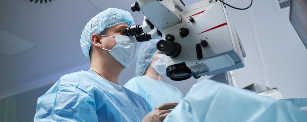
Imagine you’re in the surgeon’s chair (well, metaphorically speaking!) preparing for routine cataract surgery. You snooze through the anaesthetic, maybe occasionally sensing a gentle tug or bright light—but you’re trusting that everything will go just fine. And for the vast majority of people, it does. Cataract surgery is one of the safest and most successful operations in medicine.
But there’s one complication, rare yet serious, that can stop everything in its tracks: suprachoroidal haemorrhage. That’s the sudden bleeding inside your eye—specifically in the space between your choroid and the sclera. It might sound obscure, but if it happens, it’s an emergency, and every second counts to protect your sight.
I want to walk you through what you need to understand—without jargon. We’ll talk about risk factors, what your surgeon does immediately, and what recovery can look like if this complication does occur. Let’s dive in.
What Exactly Is a Suprachoroidal Haemorrhage?
Okay, let’s get the medical term broken down simply:
- Choroid: a layer rich in blood vessels, sandwiched between the retina (inner layer) and the sclera (tough white outer layer of the eye).
- Suprachoroidal space: the potential gap between the choroid and sclera.
- Haemorrhage: bleeding.
So a suprachoroidal haemorrhage (SCH) means there’s bleeding into that space. It can be:
- Expulsive (acute)—a sudden and dramatic event that might happen during surgery.
- Delayed (post-operative)—bleeding that occurs within hours to days after the operation.
Why does this matter? Because the eye is a closed space. If bleeding occurs, pressure builds up. That pressure can displace structures inside the eye, compress the optic nerve, or cause the choroid to “kiss” the retina—leading to rapid vision loss if not dealt with urgently.
Why It’s So Rare, Yet So Serious

Cataract surgery is extremely common—some 4–5 million in the UK every year—and most go brilliantly. Suprachoroidal haemorrhage happens in about 0.04–0.13% of cases Surgically speaking, that’s rare. Corrective action often starts within seconds of detection, and with modern surgical techniques and emergency protocols, vision loss can be prevented or minimised.
Still, because it is so rare, when it does happen, it tends to catch your attention…and rightly so. Even for surgeons, it’s a high-stakes situation that they train for. Your surgeon will know what to do, but understanding the process can help you feel more confident and prepared.
Who’s at Greater Risk?
Let’s talk about the factors that increase the chance of SCH—most of these can’t be changed on the day, but knowing them helps you and your surgeon be alert.
- High blood pressure (hypertension): Particularly if poorly controlled. High pressure can predispose weaker blood vessels in the eyeball to burst under stress.
- Age and frailty: Older patients often have more delicate tissues.
- Glaucoma or other pre-existing eye conditions: These change internal pressure dynamics in the eye.
- Myopia (high nearsightedness): Longer eyeballs can stretch blood vessels thinner.
- Use of anticoagulants (warfarin, DOACs): These are blood thinners that increase bleeding risk—though surgeons often manage these carefully.
- Previous intraocular surgery or trauma: Scarring or altered anatomy can play a part.
- Intra-operative fluctuations in eye pressure: Rapid drops in pressure—for example, when the surgeon opens the eye or injects fluid—can trigger tiny vessels to rupture.
Your surgeon will review these before your operation. Some factors can be optimised—for example, better blood pressure control—while others just mean they’ll be extra vigilant during and after surgery.
Signs That Something’s Happening
Let’s say the dreaded moment arises. What might your surgeon notice or feel?
- Rapid, unexplained increase in intraocular pressure (IOP)—the eye may feel rock solid when the surgeon palpates or irrigates.
- Loss of red reflex—that bright glow you sometimes notice during surgery. It may dull or vanish altogether.
- Sudden shallowing of the anterior chamber—the front part of the eye collapses suddenly.
- Expulsive bleeding—you might see blood push the iris forward or see it ooze into the pupil or surgical wound.
- No sign of bleeding but ongoing suspicion—this is the delayed type; may present hours later with pain, vision drop, or a firm eye.
If any of these show up, the team switches into high gear immediately.
Emergency Response During Surgery

Although rare, most surgeons rehearse their response to this every year. What might that include?
- Stop all manipulation—no more drilling or phaco-emulsification.
- Close the wound securely—to keep blood from escaping and protect the eye’s integrity.
- Discontinue irrigation-vacuum systems—to stabilise pressure inside the eye.
- Inject viscoelastic or gas—to tamponade and protect internal structures from further collapse or displacement.
- Suture swiftly and tightly, often with a very small gauge (fine stitch).
- Patch the eye or place a shield and apply gentle pressure—this helps compress the globe and limit further bleeding.
- Immediate imaging—ultrasound to assess bleeding pattern and clotting.
- Call for help—senior surgeons or vitreoretinal teams help plan definitive management.
This is about damage limitation—preventing an irreversible expulsive event and preserving what vision remains. Time is critical.
Post-operative Management
Assuming the vessel has clotted, the wound closed well, and the pressure is stable, you’re not out of the woods yet—but the immediate crisis has passed. Here’s what often happens next:
- Hospital observation—frequent IOP checks, pupil dilation, pain monitoring.
- Oral and topical steroids—to calm inflammation and reduce secondary damage.
- Intraocular pressure control—medications or even injections to keep pressure in check.
- Bed rest with head elevation—gravity helps slow bleeding and aids resorption.
- Repeat ultrasound scans—to check the clot’s size and watch for “kissing choroidals” (when the choroid folds and touches the retina, risking retinal damage).
- Drainage surgery (if needed)—once the clot shrinks (often 7–14 days later), the surgeon may carefully drain it to relieve pressure or enable retinal reattachment.
- Long-term follow up—monitor for complications like glaucoma, retinal detachment, or permanent vision loss.
Recovery is slow. It can take weeks to months for vision to stabilise.
What Does Recovery Look Like?
Every case’s different, but here’s a general idea of what to expect:
- In hospital—eye will feel bruised, pressure may fluctuate, vision may be nil or fuzzy shadows. But controlled and observed.
- Early weeks—gradual fading of pain, swelling settles, vision returns in flashes or shapes. Meds are tapered as appropriate.
- Mid-term (1 to 3 months)—vision improvement continues gradually. Some people regain nearly full acuity; others have some permanent reduction.
- Long-term—monitoring for secondary glaucoma, peripheral field changes, or retinal problems. Rehabilitation (like visual aids) may be offered if needed.
- Emotional support—this kind of complication can be scary. Talk therapy or cataract support groups can be invaluable.
Some individuals go on to have successful second-eye surgery (if applicable), once everything’s settled and healed.
Settling Nervous Minds: What You Can Do Before and After
- Talk openly with your surgeon—ask about their experience, what risk factors you might have, and how they prepare for emergencies.
- Manage your health—keep your blood pressure and any other conditions well-controlled before surgery.
- Ask about medications—are you on blood thinners, aspirin, or other agents? Your surgeon needs to plan around them.
- Set realistic expectations—even perfect surgery can’t guarantee perfect recovery. Your surgeon should explain worst-case scenarios, even if they’re very unlikely.
- Get emotional support—a trusted friend, family, or a counsellor can help if complications arise.
- Follow post-op instructions closely—take medications, don’t bend or lift heavy objects, shave or keep the eye shielded, and attend all your follow-up visits.
Looking Ahead: Vision Preservation Is Still Possible
Here’s the important takeaway: suprachoroidal haemorrhage is indeed one of the most serious complications, but with prompt recognition and expert management, surgeons often preserve meaningful vision—especially if treated calmly and without delay.
You might still recover better than you expect, though it may take longer than usual. And your surgical team is trained for this scenario. If something does go awry, they’ll act fast—every decision is geared toward protecting your sight and supporting your recovery.
Frequently Asked Questions (FAQs)
Here are ten common questions people might ask about suprachoroidal haemorrhage during cataract surgery—with thoughtful, one-paragraph answers.
1. What is the difference between expulsive and delayed suprachoroidal haemorrhage?
Answer: “Expulsive” refers to sudden, intra-operative bleeding that causes immediate pressure spikes and structural collapse—requiring the surgeon to halt, close, and tamponade the eye immediately. “Delayed” develops hours or days post-surgery and tends to present with symptoms like pain, vision drop, or increased eye firmness. Both need rapid diagnosis, but delayed cases may allow slightly more time to plan intervention, including drainage if needed.
2. Can suprachoroidal haemorrhage occur even if the surgery goes smoothly?
Absolutely. Although rare, it can occur unexpectedly, even in well-controlled surgical settings. Sharp fluctuations in eye pressure, individual weaknesses in blood vessel walls, or unseen fragility can all play a role. That’s why surgeons include it in pre-operative risk assessments and always monitor vigilantly—even during straightforward cases.
3. What are the initial signs during surgery that alert the surgeon to this complication?
Surgeons may notice a sudden loss of the red reflex (the glowing red light in the pupil), an unusually firm eye on palpation, unexpected blood appearing at the incision, or collapse of the eye’s anterior chamber. Any of these red flags triggers a rapid shutdown of activity and immediate stabilisation steps, with patient safety and vision preservation as the guiding priorities.
4. What immediate steps do surgeons take if bleeding occurs?
They stop all intra-ocular activity, close the wound tightly, use viscoelastic or gas to stabilise pressure, suture carefully, and patch or shield the eye. Imaging—such as ultrasound—is conducted urgently to assess bleeding. If needed, they may also call on retina specialists for help and plan follow-up drainage once the bleeding has settled.
5. What does hospital recovery look like after a suprachoroidal haemorrhage?
You’ll be monitored closely, with frequent checks of intra-ocular pressure, inflammation levels, and pain. Steroids—both topical and oral—help reduce swelling, while pressure-lowering medications guard against glaucoma. Elevated bed rest and head positioning aid healing, and repeat ultrasound exams track recovery. The immediate goal is stabilisation, and then gradually guiding vision back toward normal.
6. How long does it take for vision to improve after this complication?
Recovery can take weeks to months. In the first days you might see only shadows or shapes, slowly improving as swelling and clotting subside. Mid-term improvements—between 4 to 12 weeks—are common, though complete recovery depends on how promptly the bleeding was managed and whether secondary complications occurred.
7. Is further surgery often needed after a suprachoroidal bleed?
Sometimes. Once the initial bleed stabilises—often after 7–14 days—a surgeon may drain the clot to relieve sustained pressure or reattach the retina if it’s become elevated. Whether drainage is needed depends on follow-up imaging, IOP levels, and visual signs indicating whether the eye is healing naturally or still compromised.
8. Will I need extra medications long-term if this happens?
Possibly. If inflammation, raised pressure, or retinal damage persists, you may continue with topical steroids or pressure-lowering drops. In rare cases, you might need surgical implants for glaucoma or retinal repair. Long-term treatment is tailored to your individual healing trajectory and specific complications.
9. Can the second eye still be operated on after I recover?
Yes—often. Once the first eye has healed well, surgeons can assess risk for the second eye. If there are controllable factors—like blood pressure or anticoagulant medication—they’ll optimise before proceeding. Many people go on to have successful second-eye surgery, sometimes with additional precautions in place.
10. How can I prepare mentally for this possible complication?
Open communication is key. Ask your surgeon about their experience and emergency protocols. Know that it’s very rare, and should it occur, the team is trained and ready. Consider having emotional or counselling support in place ahead of surgery. Recovery can feel uncertain, but you’re not alone—and proactive understanding gives you power if anything does go wrong.
Final Thoughts
Suprachoroidal haemorrhage during cataract surgery is undeniably frightening—but rare. The overwhelming majority of operations succeed beautifully. And if you must face this complication, expert surgical intervention, careful post-operative support, and time can still preserve your vision. Keep asking questions, stay informed, and trust that your surgical team is prepared.
- Chu, T. G. & Green, R. L. (1999). Suprachoroidal hemorrhage. Survey of Ophthalmology, 43(6), pp. 471–486. Available at: https://www.sciencedirect.com/science/article/pii/S0039625799000375 (accessed 4 September 2025)
- Ling, R., Kamalarajah, S., Cole, M., James, C. & Shaw, S. (2004). Suprachoroidal haemorrhage complicating cataract surgery in the UK: a case-control study of risk factors. British Journal of Ophthalmology, 88(4), pp. 474–477. DOI: 10.1136/bjo.2003.026179. Available via PubMed: https://pubmed.ncbi.nlm.nih.gov/15031158 (accessed 4 September 2025)
- Ling, R., Cole, M., James, C., Kamalarajah, S., Foot, B. & Shaw, S. (2004). Suprachoroidal haemorrhage complicating cataract surgery in the UK: epidemiology, clinical features, management, and outcomes. British Journal of Ophthalmology, 88(4), pp. 478–480. DOI: 10.1136/bjo.2003.026138. Available via PubMed: https://pubmed.ncbi.nlm.nih.gov/15031159 (accessed 4 September 2025)

