")

Cataract surgery is one of the most commonly performed eye procedures in the world. But if you’ve previously had a retinal vein occlusion (RVO), things get more complicated. Your eye isn’t quite the same, and neither is your surgical risk. The retina—especially the macula—may already be compromised. You may have a history of swelling or fragile blood vessels. That means your surgeon needs to approach the procedure with much more caution, particularly around timing, technique, and postoperative care.
In this article, we’ll walk through the major considerations involved when planning cataract surgery in an eye that has previously experienced a branch or central retinal vein occlusion. We’ll unpack the implications of macular oedema, how anti-VEGF treatments can interact with the cataract timeline, and what strategies can help preserve your visual outcome.
Understanding Retinal Vein Occlusion: A Quick Recap
Retinal vein occlusion happens when one of the veins draining blood from the retina becomes blocked. This can be either:
- Branch Retinal Vein Occlusion (BRVO) – affecting part of the retina
- Central Retinal Vein Occlusion (CRVO) – affecting the whole retina
When blood flow is disrupted, it leads to increased pressure, swelling (especially at the macula), haemorrhages, and sometimes permanent damage to the retinal tissue. The longer the fluid and poor perfusion persist, the higher the risk of irreversible vision loss. Even with resolution of the acute phase, the eye may be left more fragile than before.
For patients needing cataract surgery after RVO, the challenge is clear: the same inflammation and fluid that compromise retinal health may also influence how the eye heals after surgery—and whether you get the visual improvement you’re hoping for.
Why Cataracts Often Follow RVO
You may wonder—why do cataracts often appear after RVO? There are a few reasons for this:
- Steroid Treatment: Many patients with RVO receive intravitreal steroids like dexamethasone implants to reduce macular oedema. While effective, steroids can accelerate cataract formation, especially posterior subcapsular cataracts.
- Age and Vascular Risk: Both RVO and cataracts become more common with age and are linked to similar risk factors like hypertension and diabetes.
- Oxidative Stress: RVO increases oxidative stress in the retina, and cataractogenesis is partly driven by oxidative damage to the lens.
So if you’ve had an RVO, you’re more likely to develop cataracts—and sooner. But the need for surgery must be carefully weighed against the risk of worsening macular swelling.
Timing Is Everything: When Should You Proceed with Surgery?
One of the most important questions your surgeon will ask is: Is the macular oedema stable?
Macular oedema—especially when associated with CRVO—is a major driver of poor visual outcomes. Cataract surgery, while beneficial, is also inflammatory. It can temporarily increase cytokines and VEGF levels in the eye, possibly worsening retinal swelling.
Surgeons typically follow these timing principles:
- Wait until the macula is dry: If there’s persistent oedema on OCT, surgery is usually postponed.
- Stability for 3–6 months: Ideally, macular thickness should remain stable for several months before surgery.
- Avoid overlap with acute anti-VEGF loading: Operating in the middle of frequent injections may disrupt the anti-inflammatory balance in the eye.
In some cases, surgeons and retina specialists coordinate surgery shortly after a stabilising anti-VEGF injection to reduce the risk of flare-ups.
Anti-VEGF and Cataract Surgery: Getting the Balance Right
Anti-VEGF injections—such as ranibizumab, aflibercept or bevacizumab—have revolutionised the treatment of RVO. They suppress vascular leakage and promote reabsorption of fluid. But how do they interact with cataract surgery?
Here are the key considerations:
- Pre-operative planning: A prophylactic anti-VEGF injection 1–2 weeks before surgery can reduce the risk of postoperative oedema recurrence.
- Postoperative follow-up: Some patients need ongoing injections after surgery to keep the macula dry.
- Drug access in the presence of an IOL: After cataract surgery, the vitreous becomes more liquefied and diffusion patterns can change slightly—but anti-VEGF still works effectively.
It’s important for the retina and cataract surgeons to coordinate treatment so that the transition from injection cycles to surgery is smooth and well-timed.
Choosing the Right Surgical Approach
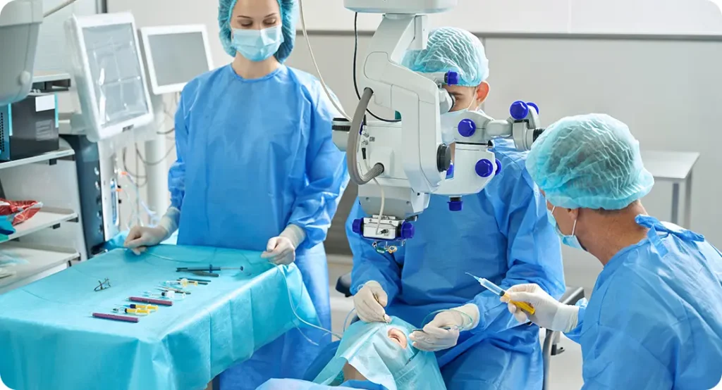
While standard phacoemulsification is generally safe in eyes with prior RVO, a few tweaks to technique can improve outcomes.
Minimising Inflammation
Postoperative inflammation can aggravate macular swelling. Surgeons may:
- Use gentle phaco parameters to reduce trauma
- Avoid prolonged energy use in the eye
- Consider pre-treatment with NSAIDs and corticosteroids
Selecting the IOL
Some surgeons prefer:
- Monofocal lenses: As they reduce visual artefacts and are more reliable in patients with compromised retinal function
- Avoidance of multifocal lenses, since these require an excellent macula to function well
Operative Anaesthesia and Monitoring
Patients with a history of retinal vein occlusion often have systemic vascular risk factors, including hypertension and diabetes. Blood pressure and cardiovascular stability during surgery should be tightly controlled, as any intraoperative spikes can affect the fragile retinal vasculature.
Postoperative Care: Watching for Red Flags
After cataract surgery, the focus shifts to protecting the retina and supporting healing. Here’s what your surgeon will monitor:
- Macular Swelling
This is the most common concern. Optical coherence tomography (OCT) is used post-op to detect any recurrence or worsening of oedema. Anti-VEGF or steroids may be reintroduced if needed. - Retinal Ischaemia
While surgery itself doesn’t worsen perfusion, any inflammatory surge could increase hypoxic stress. Surgeons keep a close eye on new capillary dropout or non-perfusion on angiography. - Intraocular Pressure (IOP)
Steroid response or previous vascular changes can lead to elevated pressure post-op. Pressure must be monitored closely, especially if a steroid implant is used. - Delayed Visual Recovery
Some patients may not experience immediate improvement in vision due to underlying retinal damage. Setting realistic expectations early on is crucial.
Predicting Visual Outcomes: What Should You Expect?
While most patients do benefit from cataract surgery, expectations must be tailored to the retinal baseline. Here’s how surgeons typically predict postoperative vision:
- Good prognosis if: the macula is dry, perfusion is intact, and pre-op vision was fair
- Guarded prognosis if: there’s longstanding oedema, macular ischaemia, or extensive vascular leakage
Surgeons often use pre-op OCT, fluorescein angiography, and visual potential testing (like potential acuity meter or laser interferometry) to estimate likely outcomes. Communication is key—patients should understand that cataract surgery won’t fix retinal damage but can still provide clearer vision if the macula is stable.
Special Considerations for CRVO vs BRVO

Not all RVOs are created equal. Here’s how the type influences surgery:
Branch Retinal Vein Occlusion (BRVO)
- Typically affects one quadrant of the retina
- Macular oedema may be more localised
- Outcomes post-cataract surgery tend to be more predictable
Central Retinal Vein Occlusion (CRVO)
- Global impact on the retina
- Higher risk of macular ischaemia and poor perfusion
- Visual outcomes are more variable
Surgeons are generally more cautious with CRVO cases and work closely with retinal specialists throughout the process.
The Role of Imaging in Pre-Op Planning
Modern imaging tools are indispensable when planning cataract surgery in eyes with a history of RVO. Key modalities include:
- OCT: To assess macular thickness, fluid, and integrity
- OCTA: To evaluate capillary dropout or non-perfusion zones
- Fluorescein Angiography (FA): For identifying leakage and ischaemia
These insights guide not only the timing of surgery but also the postoperative expectations and monitoring plan.
Coordinated Care: Why Collaboration Matters
If you’re a patient, make sure your cataract surgeon and retinal specialist are in sync. If you’re a clinician, don’t operate in isolation. A collaborative plan ensures:
- Better timing of surgery
- Appropriate anti-VEGF intervals
- Unified management of postoperative complications
Patients benefit most when care is cohesive—when everyone’s on the same page and the eye is treated holistically.
Frequently Asked Questions
1. Can cataract surgery make macular oedema worse if I’ve had a retinal vein occlusion?
Yes, it can. Cataract surgery causes a temporary increase in intraocular inflammation, which may reactivate or worsen macular oedema, particularly in eyes with a history of retinal vein occlusion (RVO). The macula, already vulnerable from past swelling or ischaemia, can respond unpredictably to the cytokine surge following surgery. This is why surgeons often delay surgery until the oedema has been stable for several months and may coordinate a pre-operative anti-VEGF injection to mitigate risk.
Even with preventive steps, there’s still a possibility of oedema returning. So, post-op OCT scans are typically scheduled within the first month to catch any recurrence early. If swelling reappears, timely treatment with anti-VEGF or corticosteroids can help maintain visual recovery.
2. Should I stop anti-VEGF injections before cataract surgery?
Not usually. In fact, anti-VEGF injections often continue before, during, and after cataract surgery in eyes with prior RVO. The injections help keep the macula dry and reduce the chance of fluid building up post-operatively. Some surgeons even administer an anti-VEGF dose 1–2 weeks before surgery to proactively suppress VEGF levels during the inflammatory response that follows cataract extraction.
The key is timing. If your injections are spaced monthly, your surgeon and retina specialist may slightly adjust the schedule to fit around the operation. Clear communication between the two teams is crucial to maintaining macular stability throughout the process.
3. Will cataract surgery restore my vision fully after a retinal vein occlusion?
It depends on how much damage your retina sustained during the vein occlusion. If the macula—the central part of the retina responsible for sharp vision—remained largely intact and dry, cataract surgery can lead to noticeable improvements. But if there was significant macular oedema or ischaemia, your visual potential may be limited.
Before surgery, your team may use a potential acuity meter (PAM) or laser interferometry to estimate how much of your vision loss is due to the cataract versus the underlying retina. This helps set realistic expectations for your visual recovery.
4. Is cataract surgery riskier if I had a central retinal vein occlusion (CRVO)?
Yes, eyes with CRVO tend to carry higher surgical risk than those with branch retinal vein occlusion (BRVO). CRVO affects the entire retina, often leads to more severe macular oedema, and is more likely to be associated with poor retinal perfusion. This means that even if the cataract is successfully removed, the retina may not support high-quality vision.
These cases require more intensive imaging and longer pre-operative observation. CRVO eyes are also more prone to postoperative complications like cystoid macular oedema or increased intraocular pressure. Surgeons often coordinate more closely with retinal specialists for these patients to ensure the best outcomes.
5. What kind of intraocular lens (IOL) is recommended in these cases?
In eyes with a history of RVO, monofocal IOLs are typically preferred. These lenses provide clear distance vision and have fewer optical side effects. Since they don’t split light between focal points like multifocal or extended depth-of-focus (EDOF) lenses do, they’re more forgiving in eyes with compromised retinal function.
Multifocal lenses are generally avoided because they can reduce contrast sensitivity and may make it harder to adapt if macular function isn’t perfect. Ultimately, lens selection is tailored to the patient’s lifestyle needs and the health of their retina.
6. How soon can I have surgery after a dexamethasone implant for macular oedema?
Ideally, surgery is timed during the window of peak steroid effect—around 4 to 6 weeks after the injection. This is when inflammation is suppressed and macular thickness is usually at its lowest. The timing helps reduce the risk of postoperative flare-ups and gives the eye the best chance at visual improvement.
If the implant is recent and the oedema is under control, your retina specialist and cataract surgeon may agree that this is the optimal moment to proceed. However, if the macula shows any sign of instability, surgery is usually postponed.
7. What happens if I delay cataract surgery too long after an RVO?
Delaying cataract surgery in an RVO-affected eye can sometimes be beneficial—especially if macular oedema needs time to stabilise. However, waiting too long can have downsides. The cataract may become hypermature, making the surgery more complex. A denser lens can also obscure OCT imaging of the retina, making it harder to monitor for oedema or ischaemia.
Functionally, patients may struggle with worsening glare, reduced contrast, and lower quality of life if surgery is delayed excessively. The decision to proceed should balance the risk of visual deterioration from cataract progression against the status of the retina.
Final Thoughts: Caution, Coordination, and Customisation
Cataract surgery in patients with a history of retinal vein occlusion isn’t routine—it’s personalised. You can’t treat these eyes like every other cataract case. You need to understand the history, review the macula carefully, and plan meticulously.
If you’re a patient, don’t rush into surgery without asking the right questions. Has your macular oedema been stable? Are your retina and cataract teams coordinating your care? And do you understand what kind of vision to expect afterward?
And if you’re a surgeon—this is a chance to do what you do best: think, adapt, and operate with precision.
For patients in the UK exploring options for private cataract surgery in complex cases like these, you can find expert care at the London Cataract Centre where we work in close partnership with retinal specialists to manage challenging scenarios with confidence.
References
- Campochiaro, P.A., 2011. Ocular neovascularisation: a valuable model system. Oncogene, 30(3), pp.254–261.
https://www.nature.com/articles/onc2010428 - Larsen, M., Waldstein, S.M., Boscia, F., Gerding, H., Silverman, D., Weichselberger, A., et al., 2016. Individualised ranibizumab treatment in patients with macular oedema due to branch retinal vein occlusion: 24-month results of the BRIGHTER study. British Journal of Ophthalmology, 100(7), pp.972–979.
- Scott, I.U., VanVeldhuisen, P.C., Oden, N.L., Ip, M.S., Blodi, B.A., Jumper, J.M., et al., 2009. SCORE Study Report 1: Baseline associations between central retinal thickness and visual acuity in patients with retinal vein occlusion. Archives of Ophthalmology, 127(9), pp.1101–1107.
https://jamanetwork.com/journals/jamaophthalmology/fullarticle/1106500 - Hykin, P., Sivaprasad, S., Prevost, A.T., Vasconcelos, J.C., Murphy, C., Kelly, J., et al., 2016. A randomised trial comparing aflibercept and bevacizumab for the treatment of macular oedema due to central retinal vein occlusion. Ophthalmology, 123(4), pp.842–850.
https://www.ncbi.nlm.nih.gov/pmc/articles/PMC4800881/ - Korobelnik, J.F., Holz, F.G., Roider, J., Ogura, Y., Simader, C., Schmidt-Erfurth, U., et al., 2014. Intravitreal aflibercept injection for macular oedema resulting from central retinal vein occlusion: one-year results from the COPERNICUS study. American Journal of Ophthalmology, 158(5), pp.1032–1038.