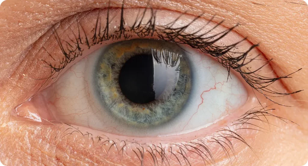
When it comes to cataract surgery, most people focus on the cloudy lens that needs replacing. But if you’re planning your own surgery—or helping someone else prepare—you might be surprised to learn that pupil size plays a far bigger role than you’d expect. From how easily your surgeon can perform the procedure, to what kind of lens you’re best suited for, and even your chances of seeing halos or glare afterwards—your pupil diameter, particularly in low light, quietly shapes the whole process.
In this article, we’ll break down the current evidence on why pupil size matters in cataract surgery. Whether you’re a patient researching your options or a clinician reviewing the latest research, this guide walks you through the essential insights, one topic at a time.
Why the Pupil Is More Than Just a Window to the Lens
The pupil isn’t just a hole that lets light into your eye—it actively changes size depending on the lighting conditions, emotional state, and age. For cataract surgery, two specific pupil behaviours are especially relevant: scotopic (dark room) and mesopic (dim room) pupil diameters.
Mesopic pupil size tends to reveal how your eyes behave in low-light but not pitch-black settings—like driving at night or walking into a dimly lit restaurant. This is the state where many visual complaints arise after surgery, especially if the wrong intraocular lens (IOL) is chosen or the optics are poorly centred.
It’s also the pupil size that might determine how well you’ll tolerate advanced IOLs like multifocal or extended depth-of-focus (EDOF) lenses. So, it’s not just a surgical technicality—it can shape your entire post-op experience.
Mesopic Pupil Diameter: A Key Metric in IOL Planning
One of the most significant advances in cataract surgery has been the availability of premium IOLs. These lenses can correct presbyopia, astigmatism, and even offer a continuous range of focus. However, they’re not ideal for everyone—and mesopic pupil size is one of the big deciding factors.
Studies suggest that a smaller mesopic pupil (typically under 3 mm) may reduce the effectiveness of multifocal lenses, particularly those that rely on ring-based designs. These lenses split light into multiple focal points. If the pupil is too small, some of these zones don’t get used, leading to reduced contrast sensitivity or fluctuating vision quality.
On the other end, a larger mesopic pupil may allow for better utilisation of all lens zones but comes with a higher risk of unwanted visual phenomena such as halos and glare. That’s why some lens manufacturers now tailor their lens recommendations based on pupil behaviour under dim light—it’s about maximising visual gain while reducing visual compromise.
Surgical Exposure and Intraoperative Considerations
For the surgeon, pupil size can make a big difference in how easily they can carry out the procedure. A well-dilated pupil gives excellent visibility of the cataract and surrounding structures, making capsulorhexis (creating the circular opening in the capsule), phacoemulsification, and lens implantation far smoother.
Smaller pupils—especially those that don’t respond well to dilation drops—pose a challenge. They increase the risk of complications such as:
- Incomplete capsulorhexis
- Iris trauma
- Prolonged surgical time
- Posterior capsule rupture
To manage this, ophthalmologists might use pupil expansion devices like Malyugin rings or iris hooks. While these tools are very effective, they do add complexity to the procedure and may affect postoperative inflammation.
Therefore, identifying a small pupil before surgery lets the team plan ahead—deciding on additional tools or even adjusting the anaesthetic strategy to ensure pupil dilation is maintained during the operation.
Dysphotopsia: When Vision Gets Annoying
Dysphotopsia refers to unwanted visual effects such as glare, halos, starbursts, or dark shadows in the peripheral vision. And yes—your pupil size is directly tied to your risk of experiencing these, especially after premium IOL implantation.
Let’s break it down:
- Positive dysphotopsia involves light phenomena (halos, glare), often seen in patients with larger pupils postoperatively, particularly under mesopic or scotopic conditions.
- Negative dysphotopsia refers to shadows or dark arcs in the temporal field of vision and may relate to light hitting the edge of the IOL-optic or the capsule.
A larger pupil in dim light means light rays from the periphery of the lens—and even from outside the optic zone—are entering the eye, leading to optical artefacts. This is especially pronounced with certain IOL materials or edge designs. If a patient has naturally large pupils, some surgeons may opt for monofocal or EDOF lenses with lower dysphotopsia profiles.
This risk needs to be discussed preoperatively, especially with patients considering night driving or those particularly sensitive to visual disturbances.
Age-Related Pupil Changes and Their Impact

As we age, the muscles responsible for pupil dilation weaken. This leads to smaller mesopic and scotopic pupil sizes, a phenomenon known as senile miosis. You might think this is a good thing—it actually can be.
Older patients often have more consistent and smaller pupil diameters in all lighting conditions. This makes them less likely to experience issues with multifocal IOL zones or peripheral light scatter. On the flip side, their smaller pupils may reduce the effectiveness of some lens technologies and limit surgical visibility.
For younger cataract patients—such as those with traumatic cataracts or high myopia—their larger pupils present both an opportunity and a challenge. They can potentially benefit from the full optics of advanced lenses but must be carefully selected to avoid high levels of dysphotopsia.
Measuring Pupil Size Accurately: Tools and Techniques
To make these decisions clinically relevant, pupil size needs to be measured objectively. Relying on visual estimates during a consultation simply won’t do.
Technologies used include:
- Infrared pupillometry – for scotopic and mesopic pupil size in standardised lighting
- Anterior segment optical coherence tomography (AS-OCT) – provides cross-sectional imaging
- Scheimpflug imaging – available on devices like the Pentacam, often used alongside topography
It’s important that these measurements are taken under controlled conditions, as stress, medications, and room lighting can all influence the results. Some clinics even test patients at multiple times of the day or simulate night-time conditions to make a more informed decision.
Implications for IOL Centration and Tilt
The size of the pupil also affects where the IOL needs to sit. Ideally, the optical centre of the IOL should align with the visual axis, not necessarily the anatomical centre of the pupil. But when the pupil dilates widely, even slight decentration or tilt becomes more obvious, especially with diffractive or aspheric lenses.
A patient with a larger pupil may notice more aberrations if the IOL isn’t perfectly aligned. This becomes a bigger issue with multifocal IOLs, which are more sensitive to these shifts compared to monofocal ones. For these individuals, precise biometry, careful surgical technique, and sometimes intraoperative aberrometry become critical to success.
Role of Pupil Dynamics in Refractive Surprise
Refractive surprise is when the final vision after cataract surgery isn’t quite what was planned. It’s not always due to poor measurements—it can also be the result of dynamic pupil changes.
If your pupil size changes significantly depending on lighting, your eye might “prefer” different focal zones at different times. This is particularly relevant with multifocal lenses that distribute light across zones. If one zone dominates under dim light but is less effective for distance, you might end up with inconsistent vision that feels off, even if the biometry was technically correct.
Understanding pupil dynamics preoperatively gives the surgeon a chance to plan more personalised solutions—perhaps using monovision, choosing an EDOF lens, or sticking with a safer monofocal option.
What About Laser-Assisted Cataract Surgery?
Femtosecond laser-assisted cataract surgery (FLACS) offers ultra-precise capsulotomies and lens fragmentation. However, pupil size still matters—perhaps even more so.
The laser systems require a minimum pupil diameter to work effectively. If the pupil is too small, suction and docking become challenging. Also, iris movement or insufficient dilation can interfere with laser beam placement, risking incomplete capsulotomies or non-uniform lens segmentation.
For patients with a history of narrow pupils or medications like alpha-blockers, FLACS might not be suitable unless mechanical dilation tools are used. So again, pupil behaviour informs surgical planning at a very early stage.
Medications and Systemic Factors That Influence Pupil Size
You’d be surprised how many medications affect pupil size. Common culprits include:
- Alpha-blockers – used for prostate issues; they can cause intraoperative floppy iris syndrome (IFIS)
- Antidepressants – may affect pupillary response
- Opiates and benzodiazepines – often lead to pinpoint pupils
- Stimulants – may enlarge pupils unpredictably
These factors should be discussed during your pre-op assessment. Sometimes, stopping or switching medications ahead of surgery can improve outcomes—but this needs to be coordinated carefully with your GP or relevant specialist.
Pupil Size and Its Role in Night-Time Visual Performance

After cataract surgery, one of the biggest concerns for many patients—especially those still driving at night—is how their new lens will perform in low-light conditions. This is where mesopic and scotopic pupil sizes start to play a crucial role in real-world visual satisfaction.
At night, the pupil naturally dilates to let in more light. But this also means it may expose the peripheral parts of the IOL—especially in multifocal or trifocal designs—leading to unwanted optical artefacts. Patients with larger pupils are particularly vulnerable to seeing halos around headlights or experiencing glare from streetlamps. While some of this can improve with time as the brain adapts, certain visual disturbances may persist if the optics don’t match the eye’s behaviour in dim lighting.
This is why some surgeons opt for monofocal lenses or newer non-diffractive EDOF lenses in patients with larger mesopic pupils who are sensitive to visual imperfections. These lenses offer a more forgiving light distribution pattern and a lower incidence of dysphotopsia—making night-time vision more stable, particularly for high-demand users.
Future Directions: Custom IOL Design Based on Pupil Profiling
We’re now entering an era of highly personalised cataract surgery, where factors like corneal topography, axial length, and even lifestyle habits help determine the best surgical plan. Pupil profiling is likely to become a central part of this personalised approach.
Several IOL manufacturers are already exploring lens technologies that adapt to pupil behaviour. For instance, some experimental designs aim to adjust their optical zones dynamically based on lighting—much like how a camera lens functions. Others are focusing on wavefront-optimised optics tailored to your mesopic pupil range, not just your refractive error.
In the clinic, artificial intelligence is also starting to play a role. Some AI tools can now simulate how a specific IOL will perform in your unique eye—including how your pupil behaves under different lighting conditions. This could soon allow patients to “preview” their postoperative vision before surgery, leading to better-informed choices and fewer regrets.
As technology evolves, the once-overlooked pupil may well become one of the most important determinants of visual success in cataract surgery.
Managing Expectations: What You Should Discuss with Your Surgeon
If you’re going in for a consultation, make sure to bring up pupil size—even if the clinic doesn’t mention it first. Ask if they’ve measured it under different lighting conditions and how it might affect the choice of lens or the surgical technique.
Here are some helpful questions you might ask:
- Is my pupil size suitable for multifocal or EDOF lenses?
- Are there any added risks for dysphotopsia in my case?
- Will you use pupil-expanding devices if needed?
- How do you plan for optimal centration in patients like me?
Being informed helps you participate actively in your care—and ensures you’re not just treated based on averages, but as an individual.
Final Thoughts
Pupil size might seem like a minor detail in the grand scheme of cataract surgery, but it can significantly shape your surgical journey and long-term outcomes. From surgical exposure and IOL performance to postoperative vision quality and the risk of visual side effects, this seemingly small factor touches nearly every aspect of your care.
So if you’re considering cataract surgery in London and want to explore the best options for your eye anatomy and lifestyle, it’s worth having a detailed pre-op assessment that includes pupil dynamics. At the London Cataract Centre, our experienced team uses advanced diagnostics to tailor every procedure to your individual needs. After all, the more your surgery is customised to you, the better your chances of seeing the world clearly—and comfortably—on the other side.
Here are five Harvard-style academic references relevant to the topic of pupil size and cataract surgery planning, with direct URLs where available:
- Holladay, J.T., 2016. Quality of vision: Essential optics for the cataract and refractive surgeon. Slack Incorporated.
- Pham, T.H., Wang, M.Y. and Porco, T.C., 2022. The effect of mesopic pupil size on the outcomes of presbyopia-correcting intraocular lenses. American Journal of Ophthalmology, 239, pp.102–110.
Available at: https://doi.org/10.1016/j.ajo.2022.02.001 - Masket, S. and Fram, N.R., 2011. Pupil size and the risk of negative dysphotopsia after intraocular lens implantation. Journal of Cataract & Refractive Surgery, 37(10), pp.1855–1859.
Available at: https://doi.org/10.1016/j.jcrs.2011.03.046 - Chang, D.F. and Campbell, J.R., 2005. Intraoperative floppy iris syndrome associated with tamsulosin. Journal of Cataract & Refractive Surgery, 31(4), pp.664–673.
Available at: https://www.jcrsjournal.org/article/S0886-3350(04)01385-6/fulltext - Rocha, K.M., Soriano, E.S. and Chalita, M.R., 2018. Pupil size and visual quality after cataract surgery with multifocal IOLs. Current Opinion in Ophthalmology, 29(1), pp.42–49.

