")

If you have pigment dispersion syndrome (PDS) and are facing cataract surgery, you might have more questions than answers. How will this condition affect the operation? Are there special risks to your iris? And what about the stability of the artificial lens that will be implanted? You’re not alone in wondering—this is something that eye surgeons need to plan for carefully. In this guide, we’ll walk you through everything you need to know about how cataract surgery is handled in eyes with PDS, the unique challenges it presents, and how the latest techniques and technologies help ensure excellent outcomes.
What Exactly Is Pigment Dispersion Syndrome?
Let’s begin with the basics. Pigment dispersion syndrome is an eye condition in which pigment granules, which normally stick to the back of your iris (the coloured part of your eye), begin to flake off. These free-floating pigment particles then circulate within the aqueous humour—the fluid inside your eye—and often settle in places they shouldn’t, such as the trabecular meshwork, which is responsible for draining fluid out of the eye.
This pigment shedding may be subtle, or it can cause enough blockage to raise intraocular pressure, leading to a form of glaucoma known as pigmentary glaucoma. Most people with PDS are diagnosed in their 20s to 40s, often after a routine eye exam reveals increased eye pressure or pigment on the corneal endothelium (a sign known as Krukenberg spindle).
Why Does PDS Matter During Cataract Surgery?
You may be thinking: “If cataract surgery is about replacing the lens, what does the iris or pigment dispersion have to do with it?” It turns out, quite a bit.
Eyes with pigment dispersion syndrome tend to have anatomical quirks that make cataract surgery more technically challenging. One of the main concerns is what’s called intraoperative iris behaviour—in other words, how the iris responds during surgery. In PDS, the iris tends to be more mobile, floppy, and prone to coming forward or billowing with fluid currents. This can increase the risk of iris trauma, incomplete dilation, or complications during lens implantation.
Intraoperative Floppy Iris Syndrome: Is It the Same Thing?
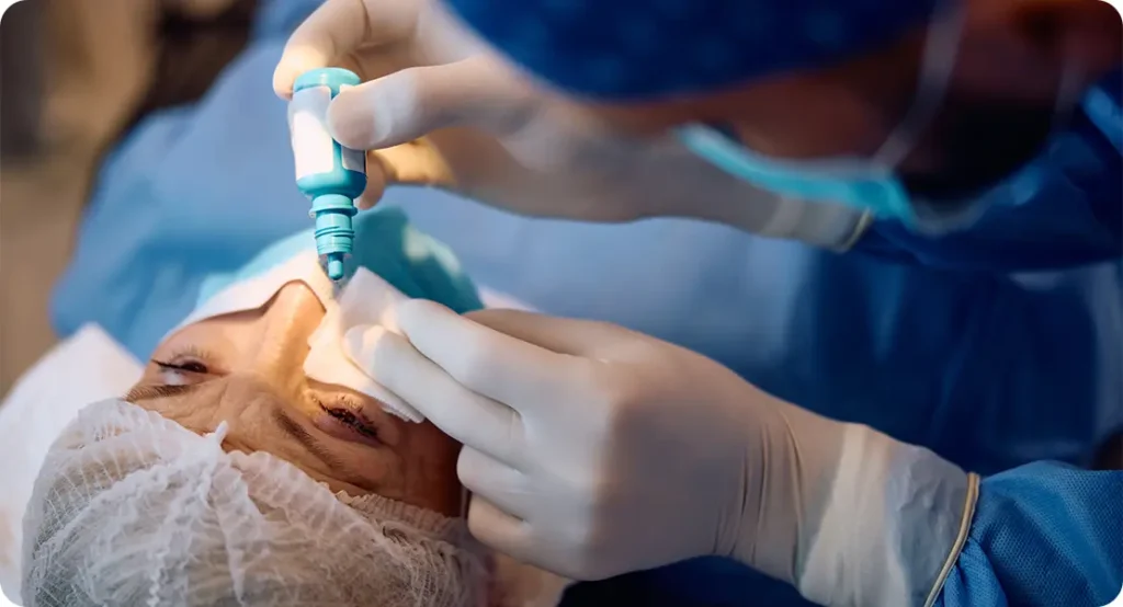
Not quite, but there is some overlap. Intraoperative floppy iris syndrome (IFIS) is most often associated with the use of certain medications like tamsulosin (Flomax), but similar floppy behaviour can also be seen in eyes with pigment dispersion syndrome due to structural differences in the iris. Surgeons therefore need to anticipate a more dynamic iris and take steps to prevent issues during phacoemulsification—the part of surgery where the cataract is emulsified and removed.
Common Intraoperative Challenges in PDS Eyes
Let’s look more closely at the actual problems that can occur when performing cataract surgery on an eye with pigment dispersion syndrome:
1. Poor Pupil Dilation
Eyes with PDS may dilate poorly or revert quickly to a smaller pupil during surgery. This limits the surgeon’s view of the cataract and increases the risk of accidental injury to the iris.
To manage this, your surgeon might use topical agents to encourage dilation and supplement them with intraoperative tools like mechanical dilators or pupil expansion devices (e.g., Malyugin rings or iris hooks).
2. Floppy Iris Behaviour
During surgery, fluid is continuously being pumped into the eye to maintain shape. In PDS, the iris can be overly mobile, fluttering or even prolapsing into surgical incisions. This complicates the procedure and can lead to bleeding, iris damage, or increased inflammation postoperatively.
Surgeons often counteract this by adjusting fluid dynamics, using low-flow techniques, and placing pupil expanders to stabilise the iris throughout the operation.
3. Zonular Weakness
While less common, some PDS patients—especially those with pigmentary glaucoma—may show signs of zonular laxity. The zonules are the delicate fibres that hold the natural lens in place, and if these are weak, the surgeon needs to be careful while removing the cataract and inserting the artificial lens.
Capsular tension rings may be used to reinforce the bag that will hold the intraocular lens (IOL), reducing the risk of future lens dislocation.
Why IOL Positioning and Centring Matters in PDS
Once the cataract is removed, an intraocular lens is implanted into your eye. In PDS, this step needs extra attention. If the lens is even slightly decentered, it could cause glare, halos, or optical distortion. More importantly, in an eye with pigment dispersion or weak zonules, a poorly placed lens might rub against the posterior iris, continuing the cycle of pigment loss and pressure spikes.
A well-centred, appropriately sized IOL placed in a stable capsular bag helps reduce these risks. Surgeons usually avoid multifocal or toric lenses in borderline cases unless they’re confident in zonular strength and IOL positioning.
Choosing the Right IOL: Does It Make a Difference?
Yes, it does. While standard monofocal lenses are generally well-tolerated, special consideration must be given when considering premium IOLs. For instance:
- Toric IOLs (for correcting astigmatism) need precise alignment. Any post-op rotation could affect vision quality and may be more likely in eyes with zonular issues.
- Multifocal IOLs (for reducing dependence on glasses) are sensitive to decentration. In a PDS eye, the added optical complexity may increase visual disturbances if stability is compromised.
- Aspheric lenses may be preferable for improved contrast sensitivity, especially if glaucoma is also present.
The takeaway: in most PDS cases, a single-piece acrylic monofocal IOL placed securely in the capsular bag is often the safest and most predictable choice.
Managing Inflammation and IOP Postoperatively
Even with a smooth surgery, eyes with pigment dispersion are more prone to postoperative inflammation. This means that you might need anti-inflammatory eye drops for a longer period. If you have pigmentary glaucoma, your intraocular pressure (IOP) may also rise temporarily after surgery, requiring a short course of pressure-lowering drops.
Some surgeons will opt for preoperative and postoperative pressure control strategies in collaboration with your glaucoma specialist, especially if your optic nerve already shows signs of damage.
When Should Cataract Surgery Be Considered in PDS?
It depends on your symptoms and your eye pressure. If your vision is significantly reduced from cataracts and you’re finding it harder to drive, read, or work, surgery is certainly justified. But if your pigment dispersion is also causing glaucoma that’s tough to control, your ophthalmologist might recommend cataract surgery earlier than usual.
That’s because removing the lens reduces its mechanical rubbing against the iris, which may help reduce pigment release and stabilise eye pressure. This benefit is particularly helpful if you’re on multiple glaucoma medications or if your IOP remains high despite treatment.
Special Considerations for Young Patients
Since PDS tends to affect younger individuals, many patients presenting for cataract surgery may still be in their 40s or 50s. This raises questions about:
- Presbyopia correction: If you’re not yet wearing reading glasses, a monofocal lens will mean you’ll need them post-op. Premium lens options should be weighed carefully.
- Long-term lens stability: Younger eyes have a longer future ahead. Ensuring the artificial lens remains stable for decades is especially important.
- Visual demands: Patients in this age group often have higher expectations and more active lifestyles, which need to be factored into surgical planning.
What About Combined Cataract and Glaucoma Surgery?
If you’ve developed pigmentary glaucoma that’s not well controlled with drops or laser treatment, your surgeon might suggest a combined procedure—cataract extraction along with a minimally invasive glaucoma surgery (MIGS) such as iStent or Hydrus.
These tiny devices help lower IOP while allowing you to potentially reduce your reliance on glaucoma medications. They can be implanted during the same operation as your cataract surgery, adding only a few extra minutes to the procedure.
Frequently Asked Questions (FAQs)
- Will cataract surgery cure my pigment dispersion syndrome?
Cataract surgery doesn’t cure pigment dispersion syndrome itself, as the underlying structural predisposition remains. However, by removing the natural lens—especially if it’s bulky and causing posterior iris chafing—the surgery may reduce further pigment shedding. In many cases, this helps to lower intraocular pressure and may even stabilise pigmentary glaucoma if it’s already present. - Is the risk of iris trauma during cataract surgery higher with PDS?
Yes, eyes with pigment dispersion syndrome often have floppy or poorly dilating irises, making them more vulnerable to trauma during surgery. Surgeons are aware of this and typically use pupil expansion devices or adjust fluid flow settings to minimise mechanical stress on the iris, helping to reduce complications like bleeding or iris prolapse. - Do I need a special kind of intraocular lens if I have PDS?
Most patients with PDS are best served with a standard monofocal intraocular lens, as it offers excellent stability and fewer risks if there’s any zonular weakness. Premium lenses such as multifocal or toric types may be considered, but only if the anatomy is suitable and the surgeon is confident about achieving a perfectly centred and stable implant. - Can PDS lead to glaucoma after cataract surgery?
Yes, pigment dispersion syndrome can still progress to pigmentary glaucoma after cataract surgery, although the risk may be somewhat reduced postoperatively. Cataract removal might lower pigment release and intraocular pressure, but patients need continued monitoring to ensure that any rise in pressure or optic nerve changes are caught early. - Is cataract surgery more painful if I have pigment dispersion syndrome?
No, the surgery itself isn’t any more painful than for other patients. The procedure is done under local anaesthesia, and while the iris may require extra handling, you shouldn’t feel discomfort. That said, you might need a slightly longer course of anti-inflammatory drops afterwards to manage inflammation more common in PDS eyes. - Can I stop my glaucoma drops after cataract surgery?
Some patients are able to reduce or stop their glaucoma drops following cataract surgery, especially if the surgery leads to a drop in intraocular pressure. However, this isn’t guaranteed, and many individuals still need ongoing medical treatment. Your ophthalmologist will monitor your pressure closely to make the right call post-op. - Will the surgeon use any special tools during my operation?
Yes, in many cases the surgeon will use tools like iris hooks or a Malyugin ring to stabilise the iris if it’s prone to floppiness or poor dilation. These devices help maintain a safe working space during surgery and reduce the risk of iris trauma, ensuring a smoother and more controlled operation. - Should I avoid cataract surgery if I have PDS?
No, there’s no reason to avoid cataract surgery solely because of PDS. In fact, if your cataract is affecting your vision or contributing to pigment dispersion, surgery may improve both vision and eye pressure control. The important part is to have the procedure done by a surgeon familiar with the nuances of operating on PDS eyes. - Can cataract surgery help lower my eye pressure?
Yes, removing the natural lens can reduce iris-lens contact, which is often the source of pigment rubbing and subsequent pressure elevation. While the pressure-lowering effect varies from person to person, some patients see enough improvement to reduce or stop their reliance on glaucoma medications after surgery. - How long is the recovery after cataract surgery with PDS?
Recovery time is generally similar to that of routine cataract surgery—usually a few weeks—but patients with PDS may experience slightly more inflammation or pressure fluctuations in the early period. Your doctor will schedule more frequent follow-ups and may tailor your drop regimen to suit your eye’s specific healing response.
Final Thoughts
If you’re living with pigment dispersion syndrome and facing cataract surgery, it’s perfectly normal to feel a bit anxious. But the good news is that with the right planning, the right team, and the right tools, surgery can go very smoothly and give you excellent visual outcomes. The key is anticipating the specific challenges your eyes may present—floppy iris behaviour, pigment instability, and lens placement issues—and addressing them with modern surgical techniques.
At the London Cataract Centre, we specialise in tailoring cataract surgery to complex eyes like yours. Whether it’s using pupil expansion devices, selecting a well-centred lens, or working closely with your glaucoma specialist, our team ensures that every step of your care is meticulously planned. If you’re ready to explore your options, we’re here to help you feel confident and informed—every step of the way.
References
- Liu L (2010) The role of the lens in pigment dispersion syndrome. Ophthalmic Surgery, Lasers & Imaging, 41(Online), e1–e4. Available at: https://pubmed.ncbi.nlm.nih.gov/21117571/ (pubmed.ncbi.nlm.nih.gov)
- Chang DF (2008) Use of the Malyugin pupil expansion device for intraoperative floppy‑iris syndrome: results in 30 consecutive cases. Journal of Cataract & Refractive Surgery, 34(5), 835–841. Available at: https://www.iogen.fi/wp-content/uploads/2011/10/2008-jcrs-malyugin-in-30-ifis-cases.pdf (iogen.fi)
- Management and outcomes of the small pupil in cataract surgery (2020). Clinical & Experimental Ophthalmology, largest observational study on iris hooks vs Malyugin rings. Available at: https://pmc.ncbi.nlm.nih.gov/articles/PMC8452752/ (pmc.ncbi.nlm.nih.gov)
- Itagaki et al. (2013) Reverse pupillary block associated with pigment dispersion syndrome after in‑the‑bag intraocular lens implantation. Journal of Cataract & Refractive Surgery, 39(12), 1925–1928. Available via DOI: https://doi.org/10.1016/j.jcrs.2013.08.020 (researchgate.net)
- Horvath KU et al. (2024) Intraoperative iris behavior during phacoemulsification maneuvers in rabbits treated with selective α1-blocker… Journal of Personalized Medicine, 14(8):840. Available at: https://doi.org/10.3390/jpm14080840 (mdpi.com)