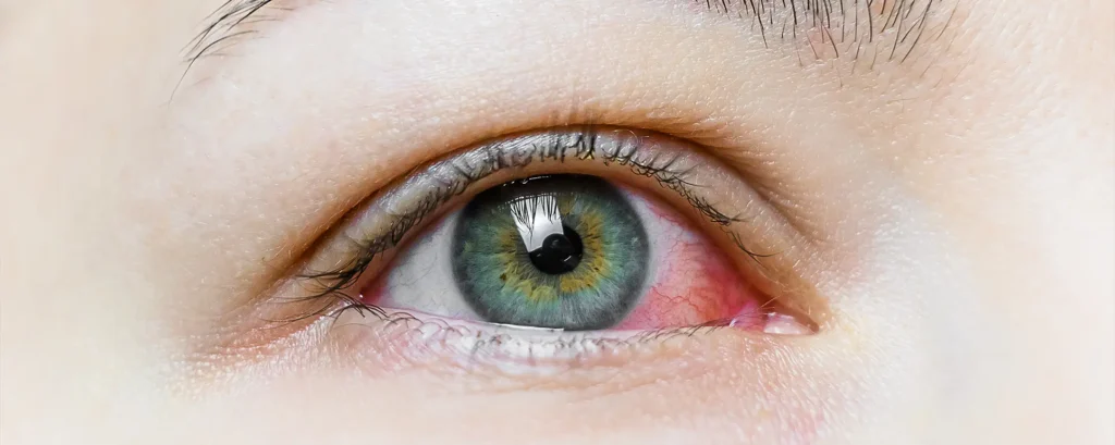
Let’s get right into it. If you’re preparing for cataract surgery—or advising someone who is—you might be surprised to hear how much the front of your eye matters. Most people focus on the lens, the cloudy part that needs removing. But what about the surface? That thin tear film, those blinking eyelids, and your meibomian glands? They quietly play a huge role in the success of surgery. Neglecting them can lead to problems no one wants: blurry results, delays in healing, and inaccurate measurements.
So yes—ocular surface health absolutely affects cataract surgery outcomes. And in this article, we’re going to explore exactly how. From the way dry eye disease (DED) skews biometric readings, to how inflammation can mess with wound healing, to the underappreciated importance of oil glands in your eyelids, we’re diving deep.
Why the Ocular Surface Matters More Than You Think
Cataract surgery may take minutes, but the preparation starts long before you lie under the microscope. The very first step? Measuring your eye. These measurements, known as biometry, guide the entire surgery. They determine which lens implant you’ll get and how well you’ll see afterwards. And here’s the thing: if your ocular surface is unstable or inflamed, those measurements can be off.
When you blink, a fresh layer of tear film coats the eye. This layer isn’t just for comfort—it’s optically crucial. It smooths out micro-irregularities and helps create a clear refractive surface. When the tear film is unstable, like in dry eye disease, your eye becomes a moving target. One reading might say you need a +20 lens. The next, a +21.5. And that can mean the difference between perfect vision and being stuck with glasses post-op.
Beyond measurements, a healthy ocular surface is essential for a smooth post-operative recovery. If your eye is inflamed to start with, it’ll be more irritable afterwards. And if your tear film isn’t working properly, you’re more prone to infection, scarring, and pain. In short, it pays to sort out the surface first.
The Tear Film Triad: Lipid, Aqueous, and Mucin Layers
Before we talk about diseases, let’s talk about what a normal tear film looks like. It’s not just salty water. In fact, it’s made up of three layers that work together:
- The outer lipid layer – Produced by your meibomian glands, this oily layer prevents your tears from evaporating too quickly.
- The middle aqueous layer – This watery portion comes from your lacrimal glands and carries oxygen and nutrients to the surface cells.
- The inner mucin layer – Secreted by goblet cells in the conjunctiva, this helps your tears stick to the eye’s surface evenly.
When any one of these layers is out of balance, the entire system wobbles. You can end up with rapid tear evaporation, inflammation, blurry vision, or all three.
Now, let’s unpack what happens when things go wrong—especially before cataract surgery.
Dry Eye Disease and Biometry: An Uncomfortable Truth

You’ve probably heard of dry eyes. They feel gritty, itchy, or watery—but they’re also a big deal when it comes to cataract planning. Dry eye disease (DED) is incredibly common in older adults, which just so happens to be the same group most likely to undergo cataract surgery.
What most people don’t realise is how dry eye impacts biometry. Instruments like optical coherence biometry and corneal topographers assume a stable surface. If your tear film is breaking up every few seconds, you’re not getting consistent readings. One scan might think your cornea is steep; another might show it as flat. And when that inconsistency sneaks into your intraocular lens (IOL) calculations, it affects your vision long-term.
Several studies have now confirmed that treating dry eye before biometry improves accuracy. Even just a few weeks of lubricating drops, anti-inflammatory agents, or lid hygiene can tighten up your measurements—and that leads to better surgical outcomes.
Meibomian Gland Dysfunction (MGD): The Hidden Saboteur
While dry eye is a broad term, a major contributor is something called meibomian gland dysfunction (MGD). These tiny oil glands, tucked inside your eyelids, are responsible for secreting the lipid layer of your tear film. When they’re clogged or inflamed, your tears evaporate faster, and the surface of your eye becomes unstable.
MGD is often missed because it can be subtle. You might not feel dry—you might just blink and notice blurry vision that clears up. But in the context of cataract surgery, MGD can wreak havoc. It not only disturbs your biometry, but it also increases your risk of postoperative irritation and slower healing.
Treating MGD is usually straightforward. Warm compresses, lid massage, omega-3 supplements, and, in some cases, in-office procedures like thermal pulsation therapy can unclog those glands and restore stability. But the key is catching it early—ideally, before any surgery is even scheduled.
Corneal Irregularities: When the Surface Goes Bumpy
An inflamed or dry ocular surface can lead to subtle corneal irregularities. These aren’t scars or scratches in the traditional sense—they’re more like tiny changes in contour or hydration that shift from moment to moment. These irregularities confuse devices like keratometers or topographers, which depend on a smooth, predictable surface.
The outcome? Your IOL power calculations can be wrong. You might also get misleading astigmatism readings. If you’re opting for a premium lens like a toric or multifocal IOL, this matters even more. Those lenses require precise alignment and power selection. A flawed surface leads to flawed assumptions—and flawed outcomes.
Addressing these issues might involve short-term use of topical steroids, cyclosporine, or even punctal plugs to reduce tear drainage and stabilise the tear film. But again, time is of the essence. The longer you wait, the harder it is to reverse those micro-changes before surgery.
Inflammation and Healing: The Domino Effect
It’s not just about how well you see after surgery—it’s also about how well you heal. An unstable or inflamed ocular surface slows epithelial regeneration, increases the risk of discomfort, and can even heighten the risk of infection. And that’s not something you want to risk when you’ve just had a tiny incision made in your eye.
Many cataract surgeons now recommend preoperative surface optimisation as standard practice. It’s a bit like prepping your skin before a major cosmetic procedure. You want things clean, calm, and hydrated. Otherwise, you’re creating the perfect storm for complications, even with the best surgical technique.
Signs You Might Have an Unstable Ocular Surface
Not sure if your surface is ready for surgery? Here are a few red flags to watch for:
- Fluctuating vision throughout the day
- Burning, stinging, or gritty sensations
- Light sensitivity or irritation in air-conditioned environments
- Redness or eyelid margin crusting
- Frequent blinking or eyelid fatigue
Even if your symptoms seem mild, it’s worth mentioning them to your eye specialist. A simple tear film assessment or meibomian gland evaluation can reveal more than you think.
Preoperative Screening: More Than Just Biometry

Modern cataract clinics are increasingly adding ocular surface assessments to their preoperative routines. This might include:
- Tear breakup time (TBUT) testing
- Schirmer’s test for tear production
- Meibography to visualise gland structure
- Fluorescein staining to detect damage
- Osmolarity testing to measure tear film stability
These aren’t just academic exercises—they provide actionable data. If your surface is compromised, your surgeon may delay biometry or surgery until it’s been optimised. It’s a short-term delay for long-term gain.
Strategies to Optimise the Surface
So, what can you do if your surgeon says your surface needs a tune-up? Here’s a common three-step approach:
1. Lid Hygiene and Warm Compresses
Your eyelids do more than just blink. They house the meibomian glands, which are responsible for secreting the oils that keep your tears from evaporating too quickly. When these glands become blocked or sluggish—a condition known as meibomian gland dysfunction (MGD)—your tear film loses its stability.
Daily lid hygiene is one of the most effective and accessible ways to manage this issue, particularly before cataract surgery. By keeping the eyelid margins clean and free of debris, you reduce inflammation, unclog the glands, and support healthier oil secretion.
Warm compresses are particularly helpful because they gently melt the meibum (oil) that may be trapped inside the glands. The warmth opens the ducts and allows for better flow, which can be followed by a light massage to express the oil.
A consistent routine—10 minutes each evening, for example—can make a noticeable difference in symptoms like fluctuating vision or discomfort. Ready-made microwaveable eye masks make the process more convenient, encouraging better compliance in the long run.
Many people don’t realise how this simple practice can have a ripple effect on surgical outcomes. A well-lubricated and stable ocular surface makes biometry more reliable and recovery more comfortable.
And unlike more invasive treatments, lid hygiene is non-pharmacological, affordable, and safe for nearly everyone. For patients preparing for cataract surgery, adding this step into their pre-op routine is one of the smartest investments they can make in their vision.
2. Artificial Tears and Anti-Inflammatory Drops
Artificial tears are the front line of defence when it comes to stabilising the ocular surface. They mimic natural tears and help maintain moisture across the cornea, reducing irritation and improving visual clarity.
Preservative-free options are generally recommended, especially for long-term use or for those with sensitive eyes. These drops can be used several times a day to keep the tear film even and stable—a small habit that makes a big difference when you’re gearing up for surgery.
In cases where dry eye is more than just occasional discomfort, anti-inflammatory drops come into play. Chronic inflammation of the ocular surface—whether due to immune response, gland dysfunction, or environmental exposure—can disrupt healing and make biometry unpredictable.

Prescription medications such as cyclosporine or lifitegrast target this inflammation directly. While they often take several weeks to show full effect, they offer long-term improvements by treating the root cause rather than just masking the symptoms.
The timing of treatment is also key. Initiating anti-inflammatory drops well ahead of surgery allows the ocular surface to recover and stabilise before the all-important preoperative scans.
Surgeons increasingly recommend starting these drops at least four to six weeks in advance if dry eye or ocular surface disease is diagnosed. When the eye is calm and well-lubricated, you’re far more likely to achieve the clarity and accuracy needed for a successful surgical outcome.
3. Supplements and Lifestyle Adjustments
Sometimes, what you put in your body matters just as much as what you put on your eyes. Omega-3 fatty acids—found in fish oils and flaxseed—are known to support meibomian gland function by improving the quality of oil secretions.
Supplements are widely available, and a daily dose can lead to noticeable improvements in both tear stability and comfort over time. It’s a subtle but powerful way to support the ocular surface, especially in those with stubborn or chronic dry eye.
Hydration is another often-overlooked factor. The tear film relies on a good balance of electrolytes and fluids to function properly. Dehydration can lead to a reduced aqueous tear layer, making eyes feel gritty or tired.
Encouraging patients to drink plenty of water and reduce caffeine or alcohol intake in the days and weeks before surgery can have a cumulative benefit on tear production. It’s easy to underestimate, but the link between systemic hydration and eye comfort is well established.
Finally, lifestyle plays a big role. Screen use, central heating, air conditioning, and long hours in dry environments all contribute to ocular surface stress. Taking regular breaks during screen time (the 20-20-20 rule), using a humidifier at home, and wearing wraparound glasses in windy conditions are simple changes that make a tangible difference.
These adjustments don’t replace medical treatments, but they do enhance and sustain their effects—creating an environment where your eyes can perform their best both before and after surgery.
Why Surgeons Are Taking This More Seriously
The days of “just removing the cataract” are long gone. Today’s cataract surgery patients expect more: sharper vision, reduced dependence on glasses, and faster recovery. With the rise of premium intraocular lenses—like multifocal, toric, or extended depth of focus (EDOF) lenses—the margin for error has shrunk dramatically. A small issue on the ocular surface can mean a big drop in visual performance, especially when you’re aiming for spectacle independence.
That’s why surgeons are now taking ocular surface health far more seriously than they did a decade ago. Many clinics now screen specifically for dry eye, MGD, and tear instability before taking any measurements.
If a problem is found, surgery might be delayed while the surface is optimised. This shift in approach reflects a growing understanding that a pristine cornea and stable tear film are just as critical to visual outcomes as surgical skill and lens choice.
Patients often think of dry eye as a comfort issue, but surgeons view it as a risk factor for poor outcomes. When the corneal surface is irregular or inflamed, it’s like trying to paint on a cracked canvas—no matter how skilled the artist, the result won’t be perfect.
Recognising this, more ophthalmologists are educating their patients early and implementing ocular surface treatments as standard pre-op protocol. It’s a new standard of care that puts long-term success at the centre of the cataract journey.
Final Thoughts
If there’s one thing to take away from all this, it’s that your ocular surface is the foundation for cataract surgery success. Skipping over it is like painting a wall without priming it first—it might look okay for a while, but cracks will show.
So if you’re planning surgery soon, speak up. Tell your ophthalmologist about any irritation, blurriness, or dryness—even if it seems minor. And if you’re a surgeon or clinician reading this, don’t ignore the surface. It might just be the most important layer of all.
If you’re considering private cataract surgery in London, addressing your ocular surface health beforehand could significantly improve your outcome. It’s a small step that makes a big difference.
References
- Trattler, W.B., Majmudar, P.A., Donnenfeld, E.D., McDonald, M.B., Stonecipher, K.G. and Goldberg, D.F., 2016. Prevalence of ocular surface disease in patients presenting for cataract surgery. Journal of Cataract and Refractive Surgery, 42(7), pp.1091–1098.
- Epitropoulos, A.T., Matossian, C., Berdy, G.J., Malhotra, R.P. and Potvin, R., 2015. Effect of tear osmolarity on repeatability of keratometry for cataract surgery planning. Journal of Cataract and Refractive Surgery, 41(8), pp.1672–1677. Available at: https://doi.org/10.1016/j.jcrs.2014.12.051 [Accessed 28 May 2025].
- Starr, C.E., Gupta, P.K., Farid, M., Hovanesian, J.A., Matossian, C. and Potvin, R., 2019. An algorithm for the preoperative diagnosis and treatment of ocular surface disorders. Ophthalmology Management, 23(6), pp.40–47.
- Matossian, C., Micucci, M.J., Song, M.A., Goyal, S. and Stonecipher, K.G., 2022. Impact of meibomian gland dysfunction treatment on ocular surface stability before cataract surgery. Clinical Ophthalmology, 16, pp.1291–1299. Available at: https://www.dovepress.com/impact-of-meibomian-gland-dysfunction-treatment-on-ocular-surface-stab-peer-reviewed-article-OPTH [Accessed 28 May 2025].
- Donnenfeld, E.D., Sheppard, J.D., Holland, E.J. and Nichamin, L.D., 2014. Managing ocular surface disease to optimize cataract surgery outcomes. EyeWorld, 19(6), pp.30–32.

