")

If you’ve ever wondered how eye surgeons manage to perform cataract operations on eyes where the view is limited—say, by corneal scars, small pupils, or dense cataracts—then you’re in the right place. We’re going to take a deep dive into one of the most powerful tools in the cataract surgeon’s modern arsenal: intraoperative OCT.
Intraoperative optical coherence tomography (OCT), particularly in its swept-source and spectral-domain forms, is no longer just a research curiosity. It’s become a vital part of complex eye surgery workflows in leading surgical centres around the world. But what exactly is it? How does it help? And why does it matter especially for the trickiest cataract cases?
Let’s break it all down.
What Is Intraoperative OCT?
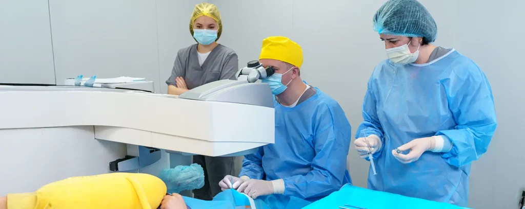
OCT, or optical coherence tomography, is essentially ultrasound—but with light instead of sound waves. It produces high-resolution, cross-sectional images of ocular structures. Traditional OCT is something you’ve probably come across in outpatient eye exams, especially if you’ve ever had a retinal scan. But intraoperative OCT takes this one step further—integrating real-time imaging into the operating theatre.
So what’s different about the intraoperative version? First, it’s live. As the surgeon operates, they can simultaneously see inside the eye in microscopic detail. Second, it’s integrated into surgical microscopes, which means there’s no need to pause the procedure or reposition the patient. It’s seamless. And in complex cataract surgeries, where precision is everything, that’s a game-changer.
Swept-Source vs Spectral-Domain: What’s the Difference?
Before we get into what intraoperative OCT can actually do, it’s worth knowing that there are two main types commonly used during surgery: spectral-domain OCT (SD-OCT) and swept-source OCT (SS-OCT). While both provide detailed images, there are a few technical differences.
Spectral-domain OCT has been around longer. It’s known for delivering high axial resolution and fast image capture speeds. However, its penetration depth can sometimes be limited in highly opaque media—such as in dense cataracts.
Swept-source OCT, on the other hand, uses a longer wavelength and has better penetration, particularly through media opacities like corneal scars or mature cataracts. That makes it especially useful in the kind of challenging surgical scenarios we’re focusing on here.
So, depending on the case and the eye’s characteristics, the surgeon might choose one over the other. But both have their strengths—and both are leaps ahead of flying blind.
Why Visualisation Is a Big Deal in Complex Cataract Cases
In standard cataract surgery, visualisation is already important. But in complex cases, it’s everything. If the surgeon can’t see the capsule clearly, or can’t judge the depth of a dislocated lens, every move becomes riskier. This is particularly true for patients with:
- Corneal scarring: This can scatter light and distort the view through the microscope.
- Dense brunescent cataracts: These block light and obscure anatomical detail.
- Posterior polar cataracts: These are fragile and prone to rupturing the posterior capsule if mishandled.
- Small pupils: These restrict access and visibility.
- Zonular weakness: This might not be visible until the surgeon starts manipulating the lens.
Intraoperative OCT offers a way around these visual challenges. By providing real-time cross-sections of the eye, it lets the surgeon see beyond the surface—and that can make all the difference.
Enhancing Capsulorhexis Accuracy
Let’s start with the capsulorhexis—the first major step in cataract surgery, where a circular opening is made in the anterior lens capsule. In most cases, this is done manually under the surgeon’s view through the microscope. But in eyes with compromised visibility, such as those with corneal scars, it can be nearly impossible to judge depth or get a perfect circle.
This is where intraoperative OCT steps in. With real-time imaging, the surgeon can ensure they’re cutting at the right depth and avoiding radial tears. In paediatric cataracts, white cataracts, or fibrotic capsules, this becomes especially helpful. The result? A safer surgery and a more predictable outcome for the patient.
Guiding Hydrodissection and Nucleus Removal
Once the capsulorhexis is complete, the surgeon typically performs hydrodissection—injecting fluid to separate the lens nucleus from the cortex. It sounds simple, but in eyes with posterior polar cataracts or compromised capsules, this manoeuvre can be risky. A little too much pressure, and the posterior capsule could rupture.
Intraoperative OCT gives immediate feedback on how the fluid wave is moving and whether the lens is properly separated. If there’s too much tension or the posterior capsule looks too thin, the surgeon can switch to a different technique—like viscodissection—to avoid a rupture.
During phacoemulsification, intraoperative OCT can also help guide the depth of sculpting, especially in dense lenses. This reduces the risk of inadvertently plunging too deep and rupturing the posterior capsule.
Managing Posterior Capsule and Vitreous Face
One of the biggest fears in complex cataract surgery is damaging the posterior capsule. It’s thin, delicate, and invisible in certain lighting conditions. If it ruptures, the lens fragments can fall into the vitreous cavity, turning a routine surgery into a much more complicated one.
Intraoperative OCT allows the surgeon to clearly visualise the capsule during critical moments. If there’s any suspicion of rupture, they can confirm it immediately and adapt their plan—whether that means switching to a posterior-assisted levitation technique or planning a pars plana vitrectomy.
OCT also lets surgeons assess the vitreous face, which becomes crucial if they need to decide whether vitreous has prolapsed into the anterior chamber. Rather than relying solely on tactile feedback or triamcinolone staining, they can see what’s happening in real-time. That’s a huge leap in surgical safety.
Ensuring Accurate IOL Placement
In standard cases, intraocular lens (IOL) placement is usually straightforward. But in eyes with weak zonules, previous trauma, or post-vitrectomy changes, getting the lens in the right place—and making sure it stays there—is much trickier.
Intraoperative OCT lets the surgeon check exactly where the haptics are landing. Are they in the sulcus or the bag? Is the optic properly centred? Is there any tilt? These might sound like technical details, but they’re key to achieving the best possible visual result—and avoiding IOL dislocation post-operatively.
In patients receiving toric or multifocal IOLs, even a slight misalignment can compromise visual quality. OCT helps fine-tune this placement for optimal outcomes.
Complex Scenarios Where Intraoperative OCT Really Shines
Let’s take a look at some real-world examples where intraoperative OCT can completely change the game:
- Post-corneal transplant patients: OCT helps assess graft-host junctions and anterior chamber stability throughout the surgery.
- Eyes with previous trauma: Intraoperative OCT reveals hidden posterior capsule defects and helps evaluate angle structures.
- Femtosecond laser-assisted surgery: OCT confirms laser cut depths, bubble layers, and capsulotomy quality.
- Anterior segment anomalies: It assists in identifying anterior segment dysgenesis or congenital structural abnormalities before making surgical decisions.
These aren’t rare cases. Any busy cataract surgeon will encounter these scenarios regularly. And in each one, intraoperative OCT provides clarity that simply wasn’t available before.
Teaching Tool and Training Aid
There’s another unsung benefit here—especially for teaching hospitals and training programmes. Intraoperative OCT gives trainees a whole new layer of insight into the anatomy and mechanics of surgery. It’s like having an x-ray machine while you’re learning to drive. Instead of just guessing where the instruments are going, you can see it—live, in real-time.
This can improve confidence, reduce errors, and accelerate the learning curve for young ophthalmologists. It also provides an opportunity for mentors to explain complex structures and responses during the actual procedure.
The Downsides and Limitations
No technology is perfect—and intraoperative OCT is no exception. There are limitations to consider.
First, the cost. These systems are expensive, both in terms of initial investment and maintenance. Not every facility can afford them, and even in places that have them, they may be reserved only for complex or high-risk cases.
Second, there’s the learning curve. Getting comfortable with interpreting OCT images during surgery takes practice. It’s a different skill set from the visual and tactile cues most surgeons rely on. But like any new technique, it becomes second nature with time.
Third, not all structures can be visualised equally well. Highly opaque media can still limit image quality, even with swept-source systems. And in some cases, motion artefacts or shadowing may reduce the utility of the images.
Still, these are trade-offs many surgeons are happy to make—especially when the benefits can mean fewer complications and better outcomes.
What the Research Says
A growing body of peer-reviewed research has highlighted the tangible benefits of using intraoperative OCT during cataract surgery, particularly in complex or high-risk cases. Numerous studies have reported that the technology significantly enhances surgical precision, which can translate to better outcomes for patients.
One of the most frequently cited advantages is the reduction in posterior capsule rupture rates—a serious complication that can arise when the surgeon cannot clearly see the capsule’s integrity. By providing real-time visualisation of delicate structures during surgery, intraoperative OCT allows for more careful handling and better decision-making, especially in eyes with pre-existing pathology.
In addition to structural safety, research has demonstrated that intraoperative OCT improves the accuracy of IOL centration and alignment. This becomes especially important when using advanced IOLs such as toric or multifocal lenses, where even slight deviations can lead to suboptimal visual results.
Several observational studies and case series have documented that when surgeons use intraoperative OCT to guide IOL placement, there is less postoperative lens tilt and fewer refractive surprises. This precision not only improves functional outcomes but also leads to higher patient satisfaction—an important metric in modern cataract practice.
Intraoperative OCT also appears to add particular value in refractive cataract surgery, where patients often expect spectacle independence and highly tailored visual correction. Research has found that OCT-guided procedures result in fewer adjustments post-surgery, such as lens repositioning or enhancement procedures. For clinics focused on delivering premium cataract care, these findings suggest that intraoperative OCT is not just a helpful tool—it’s a key component of delivering on patient expectations.
Some studies have even explored the integration of OCT imaging with digital planning platforms, adding another layer of predictive power to intraoperative decision-making.
While large-scale, long-term randomised controlled trials are still relatively limited, the general consensus among ophthalmic researchers and clinicians is increasingly positive.
Meta-analyses and reviews published in respected journals affirm the utility of intraoperative OCT across a variety of challenging surgical scenarios. What’s clear is that this isn’t a technology reserved for elite centres or cutting-edge experimental setups anymore—it’s gradually becoming an evidence-backed mainstay in cataract surgery, particularly when precision and safety are paramount.

Frequently Asked Questions (FAQs)
1. What is intraoperative OCT and how is it used in cataract surgery?
Intraoperative OCT (Optical Coherence Tomography) is an advanced imaging tool that gives surgeons a cross-sectional view of the eye during surgery. It’s particularly valuable in cataract procedures where visibility may be compromised. By offering real-time imaging through the surgical microscope, it helps the surgeon confirm structures like the lens capsule, zonules, and vitreous face—making surgery safer and more precise.
2. How does intraoperative OCT help in eyes with corneal scars or dense cataracts?
Corneal scars and dense cataracts can make it hard for surgeons to see through the microscope during surgery. Intraoperative OCT uses light waves to see beyond the cloudiness, producing detailed images of the eye’s internal structures. This clarity is crucial for safely performing steps like capsulorhexis and lens removal in otherwise high-risk eyes.
3. What is the difference between swept-source and spectral-domain intraoperative OCT?
Swept-source OCT uses longer wavelengths, giving it deeper tissue penetration—ideal for seeing through dense cataracts or opacities. Spectral-domain OCT, on the other hand, offers superior resolution but can struggle with penetration in opaque media. Both are valuable, but surgeons may prefer one over the other based on the specific challenge they’re facing.
4. Can intraoperative OCT prevent complications during surgery?
It can’t guarantee a complication-free surgery, but it certainly helps reduce the risk. By allowing the surgeon to visualise key structures during each step—especially the fragile posterior capsule or prolapsing vitreous—OCT helps in making informed decisions quickly, potentially avoiding issues like capsule rupture or incorrect IOL placement.
5. Is intraoperative OCT used in every cataract surgery?
No, it’s typically reserved for complex or high-risk cases due to its cost and the added setup time. Eyes with previous trauma, corneal scarring, or advanced cataracts are prime candidates. For routine cases, standard visualisation is usually sufficient. However, some premium surgical centres may use it more broadly to enhance precision even in straightforward operations.
Final Thoughts: Is It Worth It?
So, is intraoperative OCT the future of cataract surgery? It’s already a major part of the present. Particularly in complex cases, it’s proving its worth in theatre after theatre.
If you’re a surgeon operating on dense cataracts, scarred corneas, or eyes with unpredictable anatomy, this technology doesn’t just make things easier—it can make things safer. And for patients, that translates into better outcomes, quicker recovery, and a reduced risk of complications.
In a world where precision matters more than ever, intraoperative OCT is proving itself not as a luxury—but a necessity.
References
- Ehlers, J.P., Dupps, W.J., Kaiser, P.K., Goshe, J.M., Singh, R.P., Petkovsek, D. and Srivastava, S.K., 2014. The prospective intraoperative and perioperative ophthalmic imager–surgical assessment (PIONEER) study: 2-year results. American Journal of Ophthalmology, 158(5), pp.999–1007.e2.
- Titiyal, J.S., Kaur, M., Falera, R., Rathi, A., Kinkar, A. and Sharma, N., 2020. Intraoperative optical coherence tomography in anterior segment surgeries. Indian Journal of Ophthalmology, 68(11), pp.2381–2388. Available at: https://www.ijo.in/text.asp?2020/68/11/2381/299401
- Ehlers, J.P., Tao, Y.K., Srivastava, S.K., Hu, M., Maldonado, R.S. and Izatt, J.A., 2011. Integration of a spectral domain optical coherence tomography system into a surgical microscope for intraoperative imaging. Investigative Ophthalmology & Visual Science, 52(6), pp.3153–3159. Available at: https://iovs.arvojournals.org/article.aspx?articleid=2126523
- Hirnschall, N., Findl, O. and Menapace, R., 2013. Evaluation of intraocular lens position using intraoperative optical coherence tomography. Journal of Cataract & Refractive Surgery, 39(5), pp.645–649.
- Ehlers, J.P., Ray, R. and Srivastava, S.K., 2015. The use of intraoperative OCT in cataract surgery with coexisting ocular pathology. Current Opinion in Ophthalmology, 26(1), pp.16–22. https://doi.org/10.1097/ICU.0000000000000104