If you’ve ever had cataract surgery—or are preparing for it—you’ve probably heard about how important it is to get the intraocular lens (IOL) power just right. Choosing the correct lens is crucial for seeing clearly without glasses after surgery. That’s where intraoperative aberrometry comes into play. It’s a real-time measuring tool that’s been generating plenty of buzz in the ophthalmology world. But here’s the million-pound question: does it actually make a difference?
In this article, we’ll break it all down for you—what intraoperative aberrometry is, how it works, what the evidence says, and whether it genuinely improves the accuracy of your vision post-surgery. We’ll also explore specific scenarios where it might shine (or fall short), and give you the most up-to-date findings from studies around the globe. If you’re a surgeon, a patient, or just someone fascinated by eye tech, this one’s for you.
What Is Intraoperative Aberrometry?
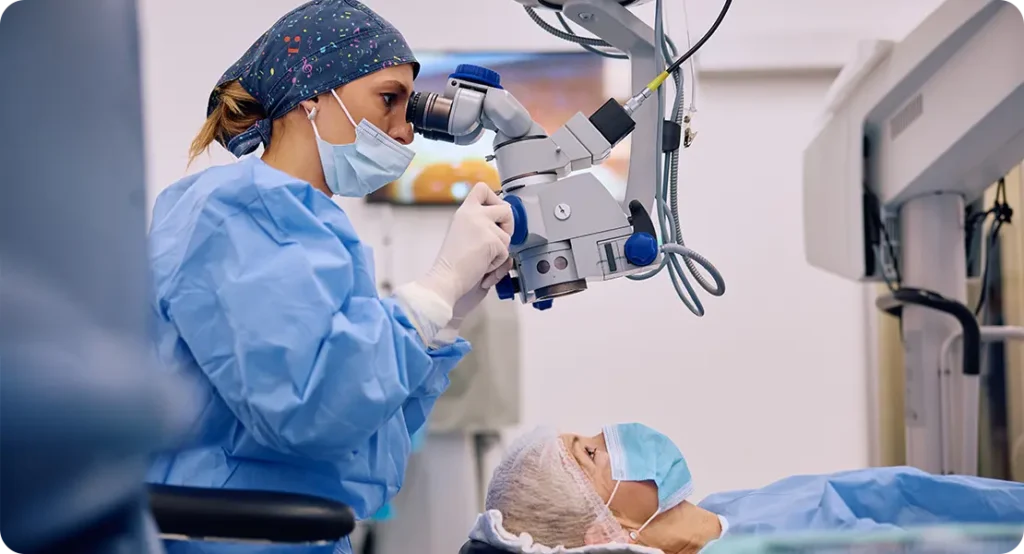
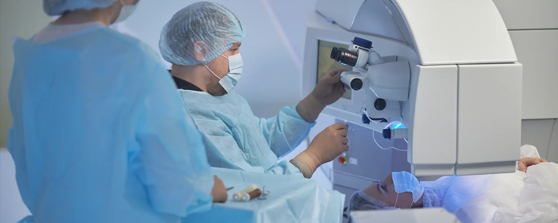
Let’s start with the basics. Intraoperative aberrometry is a technique used during cataract surgery to measure the eye’s refractive power in real time—after the cloudy natural lens has been removed but before the artificial IOL is implanted.
Rather than relying solely on preoperative measurements like axial length and corneal curvature, intraoperative aberrometry gives the surgeon a chance to fine-tune IOL selection or placement based on actual intraoperative conditions. This is particularly useful because the eye’s internal pressure and shape can change slightly once the cataract is removed. That can throw off even the most carefully calculated plans if you’re relying only on pre-op data.
The most commonly used system is called ORA (Optiwave Refractive Analysis), developed by Alcon. It uses wavefront technology—essentially bouncing light off the retina and measuring how it returns through the optical system of the eye. This helps determine the ideal IOL power and axis placement for toric lenses right then and there.
Why Might Preoperative Measurements Be Inaccurate?
You might be wondering why we even need this extra step. After all, don’t modern biometers give us precise data?
They do. But the reality is a bit more nuanced. Preoperative biometry has come a long way, especially with devices like the IOLMaster 700 and Lenstar LS 900. These can measure axial length, corneal curvature, anterior chamber depth, and lens thickness with impressive accuracy.
But they can still fall short in certain situations. For example:
- Previous refractive surgery (like LASIK or PRK) alters the corneal surface, making it tricky to estimate true corneal power.
- Dense or irregular cataracts can introduce noise and inaccuracy into pre-op scans.
- Posterior corneal astigmatism is hard to capture and can throw off toric IOL calculations.
- Lens position variability after surgery isn’t always predictable, especially in short or long eyes.
Intraoperative aberrometry aims to fill in those gaps by measuring the eye’s refractive state in its actual, altered state during surgery—something pre-op tools can’t fully do.

How ORA and Other Systems Work
The most well-known system, as mentioned, is the ORA with VerifEye+, but there are others like Holos IntraOp and the Clarity Medical System. Let’s take a closer look at how these devices actually do their job.
With ORA, the device projects wavefront light into the aphakic eye (i.e., after lens removal) and captures how that light is distorted as it passes back through the eye’s optical pathway. This wavefront data is then processed to suggest an ideal IOL power based on the real-time refractive status.
The process typically goes like this:
- The cataract is removed, and the eye is left aphakic.
- The surgeon stabilises the eye, often with a viscoelastic and an anterior chamber maintainer.
- ORA takes multiple readings, using wavefront aberrometry to assess spherical power, astigmatism, and axis.
- A suggested IOL power is displayed on a console.
- If a toric lens is being implanted, ORA can also guide precise axis alignment after the IOL is in place.
This approach isn’t completely infallible—but it offers data that’s based on the current anatomical state of the eye, rather than on projections.
What the Studies Say: Evidence for Refractive Accuracy
Alright, let’s get to the core issue: does it actually work? Is intraoperative aberrometry worth the time and cost?
A number of studies have explored this, and the results are interesting.
1. Post-Refractive Surgery Eyes
This is perhaps where intraoperative aberrometry has its strongest evidence. A 2015 study published in Ophthalmology found that in eyes with prior LASIK or PRK, ORA-guided IOL selection reduced the mean absolute error compared to conventional formulae like SRK/T and Barrett True-K. These eyes often have irregular corneal power, making traditional calculations unreliable.
In another study involving 124 eyes post-LASIK, intraoperative aberrometry produced a refractive prediction error within ±0.5 D in 78% of cases, compared to 65% using standard pre-op formulae.
Bottom line? For post-refractive eyes, intraoperative aberrometry gives a clear advantage.
2. Toric IOL Placement
Toric lenses need precise alignment to correct astigmatism. Studies suggest that ORA improves accuracy not only in choosing the correct cylinder power, but also in aligning the IOL to the correct axis. One 2021 trial found that axis misalignment dropped significantly when ORA was used compared to manual marking techniques—leading to better postoperative uncorrected distance vision.
In toric cases, ORA seems to be a valuable tool, especially when dealing with variable or subtle astigmatism.
3. Routine Cataract Cases
This is where the benefits are less clear-cut. Several studies have shown that in standard eyes with no prior surgery or astigmatism, intraoperative aberrometry offers only a marginal improvement in refractive accuracy. In some reports, the difference in mean absolute error was less than 0.1 D—clinically insignificant for most patients.
A 2019 meta-analysis covering over 1,200 eyes found that ORA improved refractive accuracy by around 10% on average, but the difference was only statistically significant in complex or atypical eyes.
So, in routine cases, the added value of aberrometry is modest at best.
Pros and Cons of Intraoperative Aberrometry
Let’s break it down simply.
Pros:
- Real-time measurement of the eye’s refractive status after lens removal.
- Particularly useful for post-refractive surgery patients.
- Helps optimise toric IOL placement—both in power and axis.
- Useful in eyes with irregular corneas or borderline biometric parameters.
- Can potentially reduce the need for IOL exchange or post-op LASIK enhancements.
Cons:
- Added cost—both for equipment and per-use charges.
- Takes additional surgical time.
- Readings can be inconsistent if the eye isn’t stable or if intraocular pressure fluctuates.
- Learning curve for accurate and reliable use.
- Marginal benefit in straightforward cases.

When Is It Most Useful?
If you’re a patient wondering whether your surgeon should use intraoperative aberrometry during your procedure, the answer really depends on your specific eye history.
Here are some situations where it’s likely to be most helpful:
- You’ve had LASIK, PRK, or RK.
- You have mild or irregular astigmatism.
- You’re getting a toric or multifocal IOL.
- You have a history of refractive surprises in the fellow eye.
- Your corneal topography is difficult to interpret.
If your eyes are average in shape and you’re receiving a standard monofocal lens, the benefits may not justify the additional cost and time. But in complex cases? It could mean the difference between perfect vision and needing glasses.
Is Intraoperative Aberrometry the Future?
Intraoperative aberrometry is not just a fancy gadget—it represents a shift in the philosophy of cataract surgery. The field is increasingly moving towards customisation, where surgeries are tailored to the unique anatomy and visual demands of each patient. Traditional IOL selection, while often accurate, still involves an element of educated guesswork. Aberrometry reduces that uncertainty by delivering data that is live, patient-specific, and responsive to intraoperative variables. As expectations rise for outcomes that are not just “acceptable” but “perfect,” tools that support personalisation are gaining prominence.
One of the key drivers of this technology’s future is integration with artificial intelligence. AI has already started to influence preoperative planning by refining IOL formulas based on massive datasets. If this predictive power can be combined with the real-time input provided by intraoperative aberrometry, we could be looking at a major leap in precision. Future systems might even suggest real-time IOL adjustments based on evolving parameters during the surgery—minimising surprises and maximising clarity post-op.
Another exciting frontier lies in the convergence of imaging technologies. Some platforms are experimenting with intraoperative optical coherence tomography (OCT) alongside aberrometry, which could dramatically improve visualisation of lens position, capsule integrity, and posterior corneal shape. This could help in real-time fine-tuning not only the IOL power but its physical placement—something that can influence visual outcomes just as much.
Ultimately, the success of any surgical technology depends on adoption, reliability, and measurable outcomes. Intraoperative aberrometry is steadily carving out a role for itself—not as a gimmick but as part of a growing toolbox for data-driven surgery. Whether it becomes the “standard” in all cataract procedures or remains a specialised tool for complex eyes will depend on how well future innovations make it more seamless, accessible, and consistently beneficial across a wider patient base.
Cost vs Benefit: Is It Worth It?
The financial equation behind intraoperative aberrometry isn’t as straightforward as you might think. On the surface, it seems like an added cost—for both surgeons and patients. There’s the capital outlay for the equipment, the maintenance, and often a per-use disposable cost. But when you dig deeper into the economics of patient satisfaction, reduced re-operations, and fewer postoperative enhancements, the numbers start to balance out, particularly in high-volume or premium practices.
For clinics that specialise in refractive cataract surgery or see a large number of post-refractive patients, the cost-benefit ratio tends to tip in favour of adopting aberrometry. These patients have high expectations and often pay out-of-pocket for premium lenses. Even small gains in refractive accuracy can mean the difference between 20/20 vision and the need for glasses—something patients are highly sensitive to when they’re paying privately. In these contexts, the return on investment can be justified by better word-of-mouth, fewer touch-up procedures, and overall improved patient outcomes.
However, in smaller practices or public health settings, it’s a tougher sell. If most surgeries are straightforward monofocal cases with minimal astigmatism, the marginal gains from intraoperative aberrometry may not offset the financial burden. Some practices choose to use the technology only in challenging cases—like irregular corneas or high-demand patients—which helps control costs while still providing access to the tool when it matters most. This selective approach can be a practical compromise in resource-constrained environments.
Patients themselves should also be part of the decision-making process. If your surgeon offers aberrometry as an add-on and you have a history of refractive surgery or you’re investing in a toric or multifocal IOL, it’s worth discussing. The additional cost may translate into a better refractive outcome, fewer surprises, and a smoother visual recovery. On the other hand, if your eyes are relatively standard and you’re happy to use glasses occasionally, the upgrade might not be necessary. Like many things in medicine, the value lies not just in the technology itself, but in how appropriately and thoughtfully it’s used.
Real-World Surgeon Opinions
Interestingly, surveys among cataract surgeons have shown mixed opinions. Some swear by intraoperative aberrometry, particularly for challenging cases. Others feel that with careful biometry and modern formulae like Barrett Universal II, they can achieve excellent results without it.
Many surgeons view it as a valuable adjunct—not a replacement—for solid preoperative planning. In fact, some use it primarily as a confirmation tool, only switching IOLs if the intraoperative reading is significantly different from their preoperative calculations.
Ultimately, like most tools in surgery, it’s only as good as the hands using it.

Final Thoughts: Does It Really Improve Accuracy?
So, where does that leave us?
Intraoperative aberrometry isn’t magic—but it’s not marketing fluff either. It has a real, evidence-backed role to play, especially in more complex eyes or for patients seeking the most accurate possible outcomes.
If you’re getting cataract surgery and fall into one of the high-risk groups for refractive surprises—say you’ve had LASIK or you’re aiming for spectacle independence—then yes, it’s worth discussing with your surgeon. It might just be the fine-tuning tool that tips the balance from “pretty good vision” to “crystal clear.”
But if you’re a routine case, getting a standard monofocal lens, and your biometry looks rock solid? The benefit may be less noticeable.
Like so much in medicine, it comes down to personalisation—and having a good conversation with your ophthalmologist about your goals, risks, and options. If you are looking to consult with an expert private cataract surgery clinic in London, you can get in touch with us here at the London Cataract Centre to discuss whether intraoperative aberrometry is right for your case.
References
- Chang, D.H.T., Breen, M. and Fischer, S., 2015. Clinical outcomes of intraoperative aberrometry in post-refractive surgery cataract patients. Ophthalmology, 122(6), pp.1090–1099. Available at: https://doi.org/10.1016/j.ophtha.2015.02.005
- Hatch, K.M., Talamo, J.H. and Krueger, R.R., 2016. Optical principles of intraoperative wavefront aberrometry. International Ophthalmology Clinics, 56(3), pp.127–141.
- Ianchulev, T., Hoffer, K.J., Yoo, S.H., Chang, D.F. and Breen, M., 2014. Intraoperative refractive biometry for predicting intraocular lens power calculation: outcomes of the prospective multicenter study of ORA system. American Journal of Ophthalmology, 158(6), pp.1164–1169.e1.
- Cionni, R.J. and Dougherty, P.J., 2018. Use of intraoperative aberrometry to improve outcomes of toric intraocular lens implantation. Journal of Cataract and Refractive Surgery, 44(6), pp.743–747. Available at: https://www.jcrsjournal.org/article/S0886-3350(18)30306-6/fulltext
- Fram, N.R., Masket, S. and Wang, L., 2017. Comparison of intraoperative aberrometry and preoperative biometry for intraocular lens power selection in post-LASIK cataract surgery. Journal of Cataract and Refractive Surgery, 43(4), pp.500–505. Available at: https://www.jcrsjournal.org/article/S0886-3350(17)30119-1/fulltext

