")

You should understand that endothelial cells are tiny, specialised cells lining the inner surface of the cornea, responsible for keeping it clear and hydrated. When considering implantable collamer lenses (ICLs), patients often worry about potential cell loss, particularly younger individuals who expect many decades of clear vision. Maintaining a healthy endothelial layer is essential, as significant cell loss can lead to corneal swelling and reduced visual quality over time.
Before surgery, I carefully measure endothelial cell density and assess corneal thickness to ensure the eye can safely accommodate an ICL. These baseline measurements are critical for patient selection and help determine whether the procedure is appropriate. Post-operative follow-up includes regular endothelial checks, which allow me to detect early changes and intervene if necessary.
Long-term studies tracking patients over five to ten years indicate that endothelial cell loss after ICL implantation is minimal. Most patients maintain healthy cell counts, and significant declines are rare when lenses are properly sized and placed. This evidence reassures patients that the procedure does not compromise corneal integrity under careful monitoring.
In my clinical experience, combining meticulous pre-operative assessment with ongoing post-operative surveillance ensures both safety and longevity of the procedure. By addressing patient concerns about endothelial health upfront, we can achieve excellent visual outcomes while preserving corneal clarity for many years.
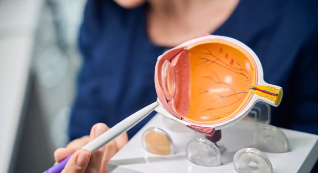
Why Endothelial Cells Are So Important
You should understand that the corneal endothelium is a single layer of specialised cells lining the inner cornea. Their critical role is to pump fluid out of the cornea to maintain clarity and prevent swelling, and unlike most cells in the body, they do not regenerate. Any permanent loss could therefore have long-term consequences for vision.
This makes endothelial health a central consideration when planning ICL surgery. Even a precisely implanted lens can lead to problems if it interferes with these delicate cells, potentially causing corneal swelling or reduced clarity over time.
I always explain to patients that ICL surgery is not only about correcting refractive errors but also about safeguarding endothelial integrity. Careful assessment and surgical technique are essential to preserve these cells, ensuring both immediate visual improvement and long-term corneal health.
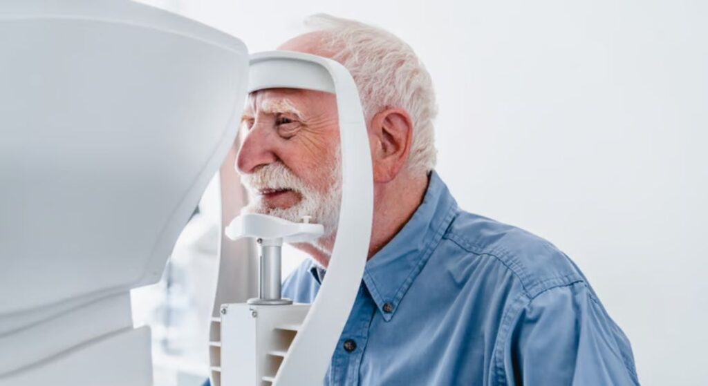
How Endothelial Cell Density Is Measured
Before surgery, I rely on specular microscopy to measure endothelial cell density (ECD) with precision. This imaging allows me to quantify cells per square millimetre and assess morphology, giving a reliable baseline before introducing an intraocular lens. In most adults, values sit between 2,500 and 3,000 cells/mm², with a gradual physiological decline of roughly 0.6% per year.
What matters in practice is not just the number, but the pattern. I assess cell size variation and hexagonality because early stress on the endothelium often shows up as morphological change before a numerical drop. This gives me an early warning system and helps determine whether a patient is a safe candidate for ICL implantation.
After surgery, I follow a structured monitoring protocol at 1 month, 6 months, 1 year, and then annually. This allows me to detect any deviation from expected cell loss trends and act early if needed. In younger patients especially, I prioritise a strong baseline and stable follow-up data, because we are planning not just for immediate outcomes but for decades of corneal health.
What Causes Endothelial Cell Loss During ICL Surgery

Endothelial cell loss during ICL surgery is usually linked to mechanical factors rather than biological intolerance. During insertion, there is a small but real risk of contact between surgical instruments, the lens, and the inner corneal surface. Even with careful handling, minimal trauma can occur, which is why controlled technique and chamber stability are essential throughout the procedure.
Post-operatively, the main concern shifts to long-term positioning of the lens inside the eye. If the vault the space between the ICL and the natural lens is too low, the lens may sit closer to the endothelium than intended. Over time, this proximity can create subtle but continuous stress on the endothelial layer, increasing the rate of cell loss beyond normal ageing.
This is exactly why I treat lens sizing as a critical decision rather than a routine step. I take detailed measurements, including anterior chamber depth and white-to-white corneal diameter, to ensure the lens sits with adequate clearance. When sizing and placement are done properly, the risk of ongoing endothelial damage is significantly reduced, and long-term corneal stability is maintained.
Long-Term Studies on Endothelial Cell Loss
We consistently see that endothelial cell behaviour after ICL implantation follows a predictable and clinically manageable pattern. The early postoperative period is where most of the measurable change occurs, and this often leads to unnecessary concern if not properly contextualised. What matters is not just the initial drop, but the long-term trajectory and stability. When we assess outcomes over years rather than months, the data becomes far more reassuring and clinically actionable.
- Initial Decline in the First Year: Most longitudinal studies report a modest endothelial cell loss of around 2–4% within the first year after surgery. This early reduction is largely attributed to surgical manipulation and the eye adapting to the implanted lens. In practice, this is expected and falls within a safe range when proper technique and case selection are followed.
- Stabilisation After Year One: After the first year, the rate of endothelial cell loss typically stabilises and begins to mirror the natural physiological decline seen in healthy, non-operated eyes. This is a critical point because it indicates that the presence of the ICL does not continue to exert ongoing stress on the endothelium. From a long-term management perspective, this stability is what allows us to consider the procedure safe.
- Preservation at 10-Year Follow-Up: Long-term data, including 10-year follow-ups, shows that most patients maintain sufficient endothelial cell density to preserve corneal clarity. This reinforces that early postoperative changes do not translate into progressive or cumulative damage. Clinically, it gives us confidence when counselling patients about durability and safety.
- Importance of Proper Sizing and Monitoring: Outcomes are highly dependent on correct lens sizing, accurate placement, and structured postoperative monitoring. When these variables are controlled, clinically significant endothelial loss is rare. In my experience, complications tend to arise only when one of these factors is compromised, not from the lens itself.
The key takeaway is that endothelial safety with ICL is not just about the first-year numbers but about long-term behaviour. When we align surgical precision with appropriate follow-up, the risk profile remains low and predictable. Patients can be reassured that early changes are expected and do not typically progress into clinically meaningful issues. Consistent monitoring ensures that any deviation is identified early and managed proactively.
Factors Influencing Long-Term Cell Loss
While long-term outcomes with ICLs are generally very good, I pay close attention to specific risk factors that can influence endothelial cell loss over time. One of the most important is anterior chamber depth. If this space is naturally shallow, the lens sits closer to the cornea, increasing the likelihood of ongoing endothelial contact or stress.
Lens vault also plays a key role, but it’s about balance rather than extremes. A vault that is too low increases proximity to the endothelium, while an excessively high vault can create abnormal pressure dynamics within the eye. Both scenarios can contribute to subtle, long-term cellular damage if not properly managed through accurate sizing and follow-up.
I also carefully screen for pre-existing corneal conditions, such as Fuchs’ dystrophy, where the endothelial layer is already compromised. In these cases, even minimal additional cell loss can become clinically significant. Combined with this, surgical technique remains a controllable factor, and I prioritise gentle insertion, stable anterior chamber control, and precise positioning to minimise trauma.
Monitoring and Follow-Up in Practice
I follow a structured monitoring protocol for every ICL patient, and I treat it as a core part of the procedure rather than an optional extra. It starts with baseline specular microscopy before surgery, giving me a clear reference point for endothelial cell density. Without that baseline, you’re effectively guessing when assessing long-term change.
After surgery, I schedule an early review at around one month to confirm lens position, vault, and overall corneal health. This is the stage where most early issues, if they occur, will show themselves. I then move to a mid-term review at six to twelve months, specifically looking for any signs of accelerated endothelial cell loss that fall outside expected physiological decline.
From that point, I maintain annual long-term follow-ups with repeat endothelial cell counts, anterior segment imaging, and vault assessment. This consistent tracking allows me to identify subtle trends before they become clinical problems. If anything looks concerning, early intervention, such as lens repositioning or exchange in rare cases, can be carried out before vision is affected.
Lens Design and Endothelial Safety
Modern ICLs, particularly the EVO ICL and EVO+ ICL models, are specifically engineered to protect the endothelium over the long term. The vault is carefully calculated for each patient, ensuring there is adequate space between the lens, the cornea, and the natural lens. This spacing is critical, as even slight miscalculations can increase the risk of endothelial contact over time.
The physical design of the lens also plays a significant role in safety. Rounded edges reduce the likelihood of mechanical irritation, while the collamer material is highly biocompatible, helping to minimise inflammation within the eye. In practice, this means you’re not only relying on surgical technique but also on a device designed to work harmoniously with ocular structures.
These refinements are reflected in long-term clinical outcomes. Studies consistently show low rates of endothelial cell loss and minimal risk of chronic damage, even in patients followed for ten years or more. When combined with accurate sizing and proper follow-up, these design features contribute to a strong safety profile and predictable long-term performance.
What Younger Patients Should Know
If you’re considering ICL surgery at a younger age, you need to think long term. You have decades of endothelial function ahead, so protecting those cells from unnecessary loss is critical. I always look for a strong baseline endothelial cell density, ideally above 2,500 cells/mm², to ensure there is enough reserve over time.
Pre-operative planning becomes even more important in this group. I assess anterior chamber depth, vault prediction, and overall corneal health carefully before recommending surgery. If any parameter looks borderline, I would rather delay or reconsider than take unnecessary risk with long-term corneal clarity.
You also need to commit to ongoing follow-up. Regular monitoring allows us to track endothelial trends and confirm that the lens remains well positioned and safe. With the right selection, precise surgery, and consistent reviews, ICLs can provide stable, high-quality vision for decades without compromising corneal health.
Practical Tips for Ensuring Long-Term Safety
Long-term success with ICL surgery is rarely about the procedure alone; it is driven by what happens before and after it. The patients who do well over five to ten years are the ones who treat this as an ongoing clinical pathway, not a one-time fix. Small decisions around surgeon selection, follow-up discipline, and symptom awareness make a disproportionate difference to outcomes. If you want predictable, stable results, these are the levers that actually matter in day-to-day practice.
- Choose an Experienced Surgeon: Surgical experience directly influences lens sizing accuracy and intraoperative handling, both of which are critical for endothelial safety. An experienced surgeon is far less likely to oversize or undersize the lens, reducing vault-related complications. In real terms, this lowers the risk of chronic endothelial stress and sets the foundation for long-term stability.
- Commit to Follow-Up: Regular postoperative reviews, particularly endothelial cell density (ECD) checks, are non-negotiable if you care about long-term outcomes. These visits allow us to track trends rather than react to late-stage problems. Patients who skip follow-ups often miss early warning signs that are otherwise simple to manage.
- Report Changes Promptly: Symptoms like blurred vision, glare, halos, or discomfort should never be ignored or delayed. Early reporting allows for quick assessment of vault, intraocular pressure, or subtle endothelial changes before they escalate. In practice, timely intervention is what prevents minor issues from becoming clinically significant.
- Maintain Ocular Health: Co-existing conditions such as dry eye or glaucoma can indirectly impact endothelial health if left unmanaged. Keeping the ocular surface stable and intraocular pressure controlled reduces additional stress on the cornea. Long-term safety is not just about the lens, but about maintaining a healthy ocular environment overall.
The reality is that ICL surgery is highly predictable when these fundamentals are followed consistently. Most complications we see are not random; they are linked to gaps in execution, monitoring, or patient behaviour. When you align surgical expertise with disciplined follow-up and proactive self-awareness, outcomes remain stable over many years. This is how you convert a good surgical result into a durable, low-risk long-term success.
Comparing ICLs to Laser Vision Correction
When you compare ICLs with laser procedures such as LASIK and SMILE, the key difference lies in how the eye is treated. Laser procedures permanently reshape the cornea, while ICLs introduce a lens inside the eye without removing tissue. This makes a significant difference when you are considering long-term structural stability and corneal preservation.
One of the major advantages of ICLs is their reversibility, as the lens can be removed or replaced if needed. I also find that for higher prescriptions, particularly significant myopia or astigmatism, ICLs tend to deliver more predictable and stable visual outcomes than laser alternatives. Because the cornea is not thinned, its natural strength is maintained, reducing the risk of long-term biomechanical issues.
From a corneal health perspective, a well-planned ICL procedure can be just as safe, and in some cases safer, than laser vision correction. The decision ultimately depends on your individual eye anatomy, prescription level, and long-term expectations. Choosing the right approach is less about technology and more about selecting what works best for your eye.
Case Examples From My Practice
I recall a patient in their early 30s with moderate myopia and astigmatism who underwent implantation with the EVO ICL. Their pre-operative endothelial cell density was 2,900 cells/mm², providing a strong baseline for long-term safety. Follow-ups at 1, 6, and 12 months showed a reduction of around 3%, which is well within expected physiological limits after surgery.
What mattered more was the long-term trend. At the 8-year mark, their endothelial cell density remained comfortably above 2,500, and the cornea stayed clear with stable, high-quality vision. Cases like this reinforce that when patient selection, lens sizing, and surgical technique are precise, endothelial health can be preserved over many years without compromise.
In another case, the patient presented with a slightly shallower anterior chamber and a lower baseline endothelial count. I adjusted the plan with careful lens sizing and aimed for a slightly higher vault to maintain safe clearance from the endothelium. At 5-year follow-up, endothelial loss was minimal, demonstrating that tailored planning is essential for maintaining long-term safety in more complex anatomical scenarios.
Long-Term Safety Data in Literature
When we step beyond individual outcomes and look at long-term published data, the pattern becomes even clearer. The safety profile of ICL is not based on short-term success but on consistent performance across extended follow-up periods. What matters here is not isolated findings, but convergence across multiple high-quality studies. When different researchers arrive at the same conclusions over a decade or more, that is where real clinical confidence comes from.
- 10-Year Endothelial Trends (Alfonso et al., 2012): Alfonso and colleagues tracked patients over a ten-year period and reported an average endothelial cell loss of around 6–8%. This level of decline closely mirrors normal physiological ageing rather than indicating procedure-related damage. In practical terms, this tells us that the ICL does not accelerate endothelial loss beyond what we would naturally expect.
- Low Risk of Corneal Decompensation (Packer, 2016): Packer’s long-term data highlights extremely low rates of corneal decompensation following ICL implantation. This is a key endpoint because it reflects real-world clinical failure rather than just numerical changes in cell density. The rarity of such complications reinforces that endothelial safety is maintained when protocols are followed correctly.
- Critical Role of Sizing, Vault, and Selection: Across multiple studies, three factors repeatedly emerge as the strongest predictors of long-term safety: accurate lens sizing, appropriate vault, and careful patient selection. These are not minor technicalities; they are the core determinants of whether the endothelium remains stable over time. When these are executed properly, the risk profile drops significantly and remains predictable.
- Consistency Across Literature and Practice: What stands out is how closely published data aligns with real-world clinical experience. The trends seen in controlled studies are replicated in day-to-day practice when the same standards are applied. This consistency is what allows us to counsel patients with confidence rather than relying on isolated outcomes.
The broader takeaway is that long-term safety with ICL is not theoretical; it is well-documented and repeatedly validated. The literature does not just suggest safety it defines the conditions under which that safety is achieved. When we adhere to those conditions, outcomes are stable, complications remain rare, and endothelial health is preserved over time. This alignment between evidence and practice is what ultimately underpins trust in the procedure.
Post-Operative Patient Education
After surgery, I make it very clear that care does not stop once the lens is in place. You need to understand that while the ICL itself is designed to be safe, maintaining corneal health is an ongoing responsibility. Regular monitoring of endothelial cell density remains essential, and I expect patients to attend annual reviews so we can track long-term trends and intervene early if needed.
I also focus on helping you recognise what is normal and what is not. Mild fluctuations in vision during the early healing phase are expected, but any persistent discomfort, blurred vision, or unusual visual changes should be reported without delay. Early action allows us to address minor issues before they develop into something more significant.
Lifestyle plays a role as well, particularly in the first few months after surgery. I advise avoiding activities that carry a higher risk of eye trauma, as protecting both the lens position and the cornea is critical during this period. With the right awareness and follow-up, you can maintain both visual quality and long-term ocular health confidently.
Integrating ICL Surgery in London Practices
If you are considering ICL surgery, choosing the right clinic is one of the most important decisions you will make. Not every provider places equal emphasis on corneal health, long-term endothelial monitoring, and precise biometric planning. I always advise focusing on centres that combine advanced diagnostics with a structured, long-term care approach rather than treating the procedure as a one-off intervention.
A high-quality clinic will assess far more than just your prescription. Detailed measurements such as anterior chamber depth, endothelial cell density, and corneal architecture are essential for accurate lens sizing and safe placement. Just as important is a clear follow-up protocol, ensuring that your eye health is monitored consistently over the years rather than only in the immediate post-operative period.
If you are exploring options, I recommend looking into ICL surgery in London where comprehensive evaluation, tailored lens selection, and ongoing monitoring are prioritised. This level of care ensures not only excellent visual outcomes but also long-term protection of corneal health and overall ocular stability.
FAQs:
1. How much endothelial cell loss is normal after ICL surgery?
You can expect a small, measurable drop in the first year, typically around 2–4%. After that, the rate stabilises and aligns closely with natural ageing, which is roughly 0.6% per year in healthy eyes.
2. Does ICL surgery cause long-term damage to the cornea?
In well-selected patients with proper lens sizing, long-term studies show no progressive damage to the cornea. The key factor is stability after the first year, which indicates the lens is not causing ongoing stress.
3. Can endothelial cells regenerate if they are lost?
No, endothelial cells do not regenerate. This is why pre-operative assessment and long-term monitoring are critical, as the body cannot replace lost cells.
4. What happens if too many endothelial cells are lost?
Significant loss can lead to corneal swelling (oedema), blurred vision, and reduced visual quality. In severe cases, additional treatment such as corneal procedures may be required, though this is rare with proper care.
5. How do you check endothelial cell density before surgery?
I use specular microscopy, which provides a precise measurement of cell density and morphology. This gives a clear baseline and helps determine whether your eye is suitable for ICL implantation.
6. How often should endothelial cells be monitored after ICL surgery?
You should follow a structured schedule: typically at 1 month, 6–12 months, and then annually. This allows us to track trends and identify any unexpected changes early.
7. Are younger patients at higher risk of endothelial cell loss?
Not necessarily higher risk, but higher responsibility. Younger patients need strong baseline cell counts and consistent long-term follow-up because they are planning for decades of vision.
8. Can incorrect lens sizing affect endothelial health?
Yes, this is one of the most important factors. Poor sizing can result in low vault, bringing the lens too close to the cornea and increasing the risk of long-term endothelial stress.
9. How do modern ICL designs improve safety?
Modern lenses like EVO ICL are designed with features such as central flow technology, smooth edges, and biocompatible materials. These reduce inflammation, improve fluid dynamics, and minimise endothelial risk.
10. Is ICL safer for the cornea than laser vision correction?
In many cases, yes especially for higher prescriptions. ICL does not remove corneal tissue, so it preserves corneal structure and biomechanics, which can be advantageous for long-term stability.
Final Thoughts: Long-Term Confidence Comes from Discipline, Not Assumptions
What I’ve seen consistently is that long-term safety with ICL is not a matter of chance, it’s the outcome of disciplined decision-making at every stage. When you combine accurate patient selection, precise lens sizing, and structured follow-up, endothelial stability becomes predictable rather than uncertain. The data supports this, but more importantly, so does real-world clinical experience over many years.
You should approach ICL as a long-term partnership with your eye care provider, not a one-time correction. The patients who maintain excellent corneal health are the ones who stay engaged with follow-up, understand their baseline metrics, and act early if something feels off. That level of awareness is what protects your vision over decades, not just the procedure itself. If you’re considering ICL Surgery in London, you can get in touch with us at London Cataract Centre.
References:
- Ruhswurm, I. et al. (2013) ‘Outcome of posterior chamber phakic intraocular lens procedure to correct myopia https://pmc.ncbi.nlm.nih.gov/articles/PMC3841253/
- Kisiel, F.B. and Gurmeet, G.J. (2024) ‘Endothelial cell loss post‑implantable collamer lens V4c: meta‑analysis https://pubmed.ncbi.nlm.nih.gov/38194352/
- Kamiya, K., Ando, W., Tsujisawa, T., Takahashi, M. and Shoji, N. (2020) ‘Effect of angle opening parameters on corneal endothelial cell density and intraocular pressure after posterior chamber phakic intraocular lens implantation’, Journal of Clinical Medicine https://www.mdpi.com/2077-0383/9/9/2704
- Chung, B. et al. (2025) ‘Ten‑year clinical outcomes of V4c implantable collamer lens implantation: longitudinal analysis of visual acuity, endothelial cell density, and vault dynamics’, Journal of the American Academy of Ophthalmology https://www.sciencedirect.com/science/article/abs/pii/S000293942400360X
- Sanders, D.R., Doney, K. and Poco, M. (2007) ‘United States Food and Drug Administration clinical trial of the implantable collamer lens for moderate to high myopia: three‑year follow‑up’ https://academic.oup.com/bmb/article-abstract/83/1/325/384535