")

When you think about cataract surgery, you probably focus on the cloudy lens, the artificial lens being implanted and the vision you hope to achieve afterwards. But what you might not realise is how much your eye’s internal structure its physical strength, elasticity and pressure dynamics can influence your final results.
These factors fall under something called eye biomechanics, a field that is becoming increasingly important in modern cataract care. Your cornea, lens capsule and even the supporting tissues inside your eye don’t just sit passively during surgery. They stretch, react, resist or reshape depending on their natural properties.
If you’ve ever wondered why some people achieve perfect clarity while others need enhancements later on, biomechanics is often the hidden reason. And today, surgeons can measure several biomechanical features before surgery, allowing them to choose the best lens implant, plan safer incisions and improve long-term refractive stability.
In this guide, I’ll walk you through how eye biomechanics affect cataract surgery outcomes. You’ll learn why corneal strength matters, how the lens capsule behaves during surgery, how your eye responds to incisions and why personalised planning can dramatically improve visual outcomes. If you’re considering cataract surgery, understanding these concepts can help you make more informed choices when discussing your options at centres such as the London Cataract Centre, where advanced biomechanical assessment is part of modern patient care.
What Are Eye Biomechanics?
Eye biomechanics refers to the way the physical structures of your eye respond to force, pressure and movement. In simple terms, it’s about how strong, flexible and stable the different parts of your eye are, especially the cornea, lens and surrounding tissues. These structures aren’t rigid they bend, stretch and adapt slightly every time you blink, focus or experience changes in eye pressure.
When we talk about biomechanics in eye care, we’re usually referring to how the cornea and lens behave under stress, how they maintain their shape and how resilient they are during procedures like laser eye surgery or cataract surgery. Understanding these mechanical properties helps surgeons plan safer, more personalised treatments and predict how your eye will heal or respond to surgical changes.
Biomechanics refers to the physical properties that determine how tissues behave under pressure or stress. In your eyes, this includes:
- Corneal rigidity how stiff or flexible your cornea is
- Corneal elasticity how well it returns to its original shape
- Lens-capsule elasticity how the membrane holding your cataract stretches
- Scleral strength how the white part of your eye supports structural forces
- Intraocular pressure interactions how the fluids inside your eye push and reshape tissue
These factors affect how your eye bends light, how stable your vision remains over time and how your eye responds during surgery. Every eye is different and that’s why biomechanics are becoming essential in tailoring cataract procedures to each person.
Why Biomechanics Matter in Cataract Surgery
Cataract surgery is a highly refined procedure, but it still relies on the natural behaviour of your tissues. Your surgeon makes tiny incisions, removes your cloudy lens and replaces it with an artificial intraocular lens (IOL). Every step is influenced by your eye’s structural properties.
Biomechanics matter because they influence:
- How your cornea changes after incisions: Your cornea isn’t just a static structure it responds to pressure, healing forces and surgical incisions. Strong, stable biomechanics mean the cornea is more likely to maintain its shape after surgery, which directly affects how light enters your eye and how your vision stabilises over time.
- How accurately your IOL sits inside your eye: The internal biomechanics of your eye influence how well an intraocular lens (IOL) positions itself after cataract or RLE surgery. A stable, well-balanced eye structure helps the lens centre properly, reducing tilt or rotation, especially in toric IOLs that correct astigmatism.
- Whether your refractive outcome matches your expectations: Good biomechanics improve the predictability of your final prescription. If your cornea or internal eye structures shift more than expected after surgery, even small changes can alter your visual outcome and leave you slightly over- or under-corrected.
- The long-term stability of your vision: Strong biomechanics reduce the chances of gradual regression or unexpected shifts in focus. This is especially important for patients with thin corneas, high prescriptions or early corneal instability, where long-term vision can drift if the eye isn’t structurally stable.
- The likelihood of needing glasses after surgery: If your cornea or IOL remains well-positioned thanks to solid biomechanics, your chances of achieving glasses-free vision are much higher. Poor stability, on the other hand, can lead to subtle distortions that may require glasses for certain tasks.
- The risk of complications: Biomechanical strength plays a major role in preventing issues like corneal ectasia, irregular astigmatism, lens rotation, and unpredictable healing. Understanding these factors helps your surgeon choose the safest and most accurate treatment for your eyes.
When surgeons understand your biomechanics ahead of time, they can plan your surgery more precisely and avoid surprises.
How Corneal Biomechanics Influence Visual Outcomes
Corneal biomechanics influence how your cornea responds to the small incisions and pressure changes during cataract surgery. If your cornea is strong and stable, it maintains its shape more predictably, helping your vision settle smoothly after the procedure. This means you’re more likely to achieve the exact visual outcome your surgeon planned.
On the other hand, a cornea that is more elastic or weaker may change shape slightly during healing, which can affect your final prescription. Measurements like corneal hysteresis, resistance and elasticity help your surgeon understand how your cornea will behave. With this information, they can plan the surgery more precisely and reduce the chance of unexpected vision fluctuations.
1. Corneal Strength Predicts Refractive Stability
A naturally weak or overly flexible cornea is more prone to subtle shape changes after surgery, and these shifts can influence your final glasses prescription. When the cornea bends or relaxes, even by a very small amount, it can create unexpected astigmatism or mild blurring. People with stronger, more stable corneas usually experience more predictable results because the tissue holds its contour during healing. This is why surgeons evaluate corneal biomechanics so carefully before cataract or refractive procedures it helps them anticipate how your eye will behave afterward.
Even though the incisions are tiny, a softer cornea can still change curvature enough to cause:
- Unexpected astigmatism: Sometimes after eye surgery, the cornea heals in a slightly uneven way, creating small amounts of astigmatism that weren’t present before. Even mild irregularity can affect sharpness, contrast and night-time clarity, which is why precise healing is so important.
- Mild blurring: A small refractive error or slight swelling in the cornea can cause soft, hazy vision. This usually improves as the eye settles, but it can also indicate that a minor enhancement may be beneficial for achieving crisp clarity.
- Refractive regression: Some eyes naturally shift a little after surgery, causing a return of mild myopia, hyperopia or astigmatism. This is known as regression and usually happens gradually. If it affects daily tasks, a touch-up procedure can correct it.
- Fluctuating vision: Your eyesight may vary from day to day due to healing changes, dryness or small alterations in corneal hydration. This tends to stabilise over time, but persistent fluctuations may suggest that the eye is still adjusting or needs further evaluation.
On the other hand, a strong cornea tends to stabilise quickly, leading to more predictable results. This is why many modern practices assess corneal biomechanics alongside standard topography.
2. Corneal Flexibility Affects How Incisions Heal
Corneal flexibility plays a major role in how these incisions settle as your eye heals. A more elastic cornea may change shape slightly during recovery, which can influence the final refractive outcome. On the other hand, a stiffer cornea tends to hold its structure more predictably but may respond differently to surgical pressure. These natural biomechanical differences help explain why two people with the same procedure can heal in slightly different ways.
The cornea’s elasticity determines how well this incision:
- Opens: During surgery, a tiny incision or flap is gently opened to access the corneal tissue underneath. This allows the laser to reshape the eye with precision while keeping the surrounding tissue undisturbed.
- Flexes: The cornea naturally flexes slightly as the laser reshapes it. This flexibility is normal and expected it helps the surface adjust to the new curvature that corrects your vision.
- Closes: Once the reshaping is complete, the flap or incision naturally closes on its own. No stitches are needed. The cornea begins to seal immediately, protecting the treated area beneath it.
- Holds its shape as it heals: As healing progresses, the cornea stabilises and maintains its new curvature. This is what gives you clearer, more focused vision in the long run. Over the following weeks and months, the corneal structure strengthens and your vision becomes increasingly stable.
A flexible cornea may swell or reshape temporarily, causing slight visual disturbance. A stiffer cornea heals more predictably. If your cornea is unusually rigid, surgeons may adjust incision location or size to avoid complications.
3. Biomechanics Influence Astigmatism Correction
If you have astigmatism, your surgeon may correct it using either a toric IOL or precise corneal incisions, and both rely heavily on good corneal biomechanics. A stable, resilient cornea holds the intended shape after these corrections, giving you clearer, more predictable vision. But if the cornea is weaker or behaves unpredictably, it may not maintain the correction as expected, leading to slight blur or residual astigmatism. This is why assessing biomechanical strength is essential it helps your surgeon choose the method that will stay stable over time.
- A toric IOL implant: A toric intraocular lens is designed specifically to correct astigmatism during cataract or refractive lens surgery. It has different powers in different meridians, allowing it to neutralise the uneven curvature of your cornea. When positioned accurately, it provides sharp, stable vision and reduces or completely removes the need for glasses.
- Limbal relaxing incisions (LRIs): LRIs are tiny, precisely placed cuts made at the edge of the cornea (the limbus). These small incisions relax the steep curve of the cornea, reducing mild astigmatism. They are quick, painless, and often used when the amount of astigmatism is low or when a toric lens isn’t necessary.
Both depend on biomechanics. A flexible cornea may respond unpredictably to LRIs. A toric lens may rotate more if the cornea doesn’t provide stable referencing forces. A stiffer cornea usually produces more precise outcomes. Surgeons rely on biomechanical data to decide which astigmatism method is safer for you.
4. Weak Biomechanics Increase Risk in Conditions Like Keratoconus
If you have keratoconus, post-LASIK ectasia or naturally thin corneas, weak biomechanics play a much larger role in how your eye responds to cataract surgery. These corneas are more prone to shape changes, so even small surgical forces can temporarily alter curvature. This doesn’t mean cataract surgery is unsafe it simply means the planning must be far more customised to ensure stability and accurate visual results.
In cases of biomechanically weak corneas, your surgeon may adjust incision placement, choose a different IOL calculation method, avoid certain premium lenses or recommend corneal strengthening treatments like cross-linking before or after surgery. Because standard measurements can be less predictable in these eyes, extra assessments such as tomography, topography and biomechanical analysis help create a safer, more personalised surgical plan.
How Biomechanics Affect IOL Power Calculations
Before your cataract surgery, your surgeon uses several key measurements to work out the precise power of the artificial lens that will replace your cloudy natural one. These include parameters such as the curvature of your cornea, the length of your eye from front to back, the depth of the anterior chamber and predictions about where the new lens will sit once implanted. Together, these form the foundation of accurate IOL calculations and help ensure clear, stable vision after surgery.
However, your corneal biomechanics also influence these numbers more than most people realise. A cornea that is unusually soft, thin or irregular may respond differently during measurement or change shape slightly after surgery, which can affect the final refractive outcome. This is why surgeons consider both structural strength and biomechanical behaviour when selecting the best formula and lens type for your eyes.
Here’s how:
1. Corneal Elasticity Affects Keratometry: Keratometry the measurement of your corneal curvature works best when the cornea has stable, predictable biomechanics. But if your cornea flexes easily, the readings can shift slightly depending on factors like tear film stability, subtle pressure on the eye, or even the technique used during measurement. These small variations can affect the accuracy of the IOL power chosen for cataract surgery, sometimes leading to minor refractive surprises afterward. To minimise this, modern diagnostic machines now include biomechanical compensation, giving surgeons more reliable data and improving overall visual outcomes.
2. Biomechanics Influence Effective Lens Position (ELP): Biomechanics also play a key role in predicting the effective lens position (ELP) the expected final resting place of your IOL after cataract surgery. Even very small shifts in lens position can change your final prescription, so accurate prediction is essential. The way your lens capsule contracts, stretches or stabilises over the weeks after surgery is strongly influenced by your individual biomechanical profile. If your eye tends to shift or settle subtly during healing, forecasting the exact ELP becomes more challenging, which means your surgeon must select and calculate your IOL with extra precision to achieve the desired visual outcome.
3. Precise Biomechanical Data Helps in Selecting Advanced IOLs: Advanced IOLs such as multifocal and extended-depth-of-focus lenses depend heavily on stable biomechanics to perform at their best. These lenses split or extend light in very precise ways, so any irregularity in the cornea or lens capsule can reduce their clarity or create visual disturbances. If biomechanical tests show an unstable corneal shape, excessive capsule elasticity, a tendency toward IOL tilt, or a higher risk of long-term refractive drift, your surgeon may advise against choosing these premium lenses. In such cases, a high-quality monofocal lens is often the safer and more predictable option, giving you clearer and more consistent vision over time.
The Lens Capsule: A Critical Biomechanical Structure
The lens capsule is a thin, elastic membrane surrounding your natural lens. During cataract surgery, your cloudy lens is The lens capsule plays a surprisingly important biomechanical role during and after cataract surgery, even though it’s just a thin, elastic membrane. When your natural cloudy lens is removed, this capsule is carefully preserved because it acts as the support structure for your new intraocular lens. Its flexibility, strength and ability to maintain shape directly influence how smoothly the procedure goes and how stable your vision will be afterward.
A healthy capsule allows the surgeon to remove the old lens without stress, keeps the new IOL securely in place and helps it stay perfectly centred. If the capsule is weak or behaves unpredictably, there’s a higher chance of issues like tilt, decentration or capsule contraction over time, all of which can subtly affect your final focusing power. This is why understanding the biomechanics of the lens capsule is essential for predicting long-term refractive stability and ensuring you get the clearest vision possible after surgery.
1. Capsule Elasticity Affects Surgical Ease: A healthy, elastic capsule stretches easily during surgery. This makes lens removal smooth and reduces the chance of tearing. If your capsule is less elastic due to age or certain medical conditions, surgery requires slower, more controlled steps. An experienced surgeon can still manage this safely, but biomechanical awareness is essential.
2. Biomechanics Affect IOL Centration: The biomechanics of your eye directly influence how well your IOL stays centred within the lens capsule. If internal forces cause the lens to tilt, shift, rotate, or move forward or backward, your vision quality can be affected, sometimes creating blur or distortion. By understanding these biomechanical factors, your surgeon can select an IOL design that is more likely to remain stable and properly aligned, ensuring sharper and more consistent visual outcomes.
3. Capsule Contraction Can Influence Long-Term Results: After surgery, the capsule naturally shrinks slightly as it heals. This is called capsular contraction. If your biomechanics indicate a higher risk of contraction for example, if you have diabetes, inflammation or pseudoexfoliation your lens choice may be adjusted. A more rigid lens or a specialised haptic design can help maintain long-term alignment.
Biomechanics and Incision Planning
The way your cornea and surrounding tissues respond to surgical incisions is closely tied to their biomechanical properties. Factors like corneal rigidity, elasticity, and resistance to deformation determine how well the eye maintains its shape during and after the procedure. A stronger, more stable cornea can tolerate incisions with minimal impact on vision, while weaker biomechanics may require careful planning to avoid distortion.
Surgeons use this understanding to decide the size, location, and orientation of each incision. By tailoring these decisions to your eye’s specific biomechanical profile, they minimise postoperative astigmatism, maintain structural integrity, and optimise how the new lens sits, which ultimately contributes to sharper, more predictable visual outcomes.
1. Location of Incisions: The location of your corneal incisions is carefully planned according to your cornea’s biomechanics, not just its surface shape. If the cornea is flexible or irregular, placing an incision in the wrong spot can induce astigmatism, create glare or visual distortions, or lead to unpredictable healing. By using biomechanical maps, surgeons can select incision sites that minimise these risks and promote stable, high-quality visual outcomes.
2. Size of Incisions: The size of corneal incisions is influenced by both the type of IOL and your eye’s biomechanical properties. While smaller incisions are generally less disruptive, certain lenses require a slightly larger entry point. Biomechanical analysis helps your surgeon decide whether a micro-incision will remain stable, whether a larger incision is safer, and if self-sealing is likely to be reliable, ensuring the safest possible outcome for your vision.
3. Wound Sealing Depends on Tissue Strength: Wound sealing after cataract surgery depends heavily on corneal tissue strength. If the cornea is weak or thin, your surgeon may adjust the incision angle, use extra hydration at the wound, or occasionally place sutures to ensure proper closure. In contrast, a strong, healthy cornea typically seals effortlessly on its own, reducing healing time and the risk of complications.
Assessing corneal biomechanics before surgery helps your surgeon anticipate how your incisions will heal, reducing the risk of unexpected complications. By understanding tissue strength and flexibility, they can tailor incision placement, size, and technique to ensure smooth, predictable wound closure and optimal visual outcomes.
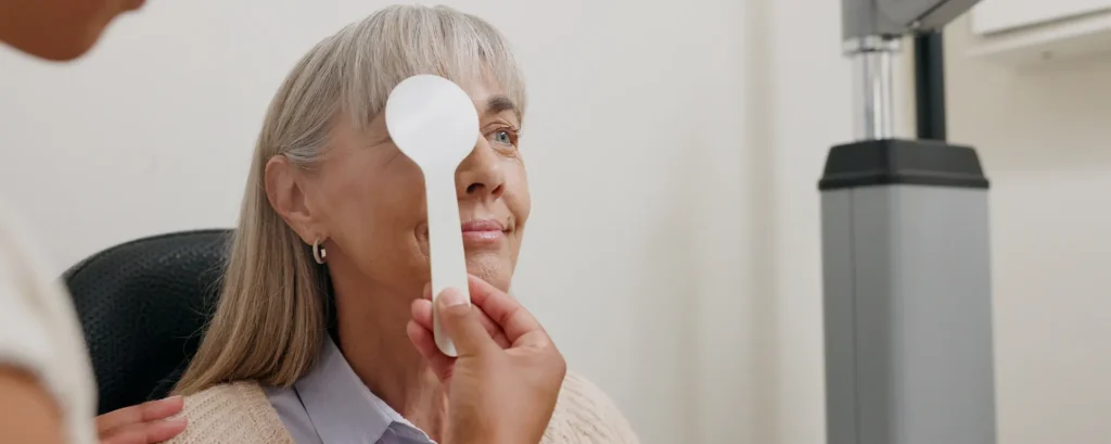
How Surgeons Measure Eye Biomechanics
Surgeons now have advanced tools to assess corneal biomechanics before surgery. Devices like the Ocular Response Analyzer (ORA) and Corvis ST use air pulses or high-speed imaging to measure corneal hysteresis, elasticity, and resistance. These tests help quantify how the cornea absorbs shock, bends under pressure, and maintains its shape.
By understanding these measurements, surgeons can tailor surgical plans more precisely. This ensures safer incisions, more stable intraocular lens placement, and a lower risk of postoperative complications such as astigmatism or lens decentration, ultimately improving visual outcomes.
Common tools include:
1. Corvis ST (Dynamic Biomechanical Analysis): The Corvis ST uses a gentle air puff and a high-speed camera to track how your cornea reacts in real time. It measures bending response, stiffness, velocity of movement, hysteresis, and pressure resistance. This detailed profile helps surgeons understand your cornea’s strength and flexibility, which is crucial for predicting how it will behave after laser surgery or other procedures.
2. Ocular Response Analyser (ORA): The ORA measures two key parameters: corneal hysteresis and corneal resistance factor. These values indicate how well your cornea absorbs and dissipates energy. This information is especially important for glaucoma patients, people with thin corneas, or anyone at risk of corneal weakening, helping surgeons assess safety before surgery.
3. Scheimpflug Tomography: Scheimpflug tomography provides a three-dimensional map of your cornea, traditionally used for curvature and shape analysis. Modern systems also evaluate depth-related stiffness patterns, giving insights into corneal biomechanics. This helps detect early signs of weakening and supports surgical planning for safe laser treatment.
4. Optical Coherence Tomography (OCT): OCT provides high-resolution images of the anterior segment, including the cornea and lens. It can detect subtle biomechanical behaviours, like tissue elasticity and capsule thickness, which influence surgical outcomes. Surgeons use this to refine treatment plans and ensure precise, stable results.
This is especially useful for surgical planning.
Who Benefits Most from Biomechanical Assessment?
Biomechanical evaluation is beneficial for all cataract patients, but it is particularly important for those with specific risk factors. Patients with thin or irregular corneas, high prescriptions, or a history of refractive surgery gain crucial insights, as their eyes are more prone to unpredictable healing or refractive changes. Those considering advanced IOLs, such as multifocal or extended-depth-of-focus lenses, also benefit, since biomechanics guide lens selection and placement. Additionally, anyone with prior corneal disease or trauma can achieve more stable and predictable long-term visual outcomes with this assessment.
- Have astigmatism
- Have had previous laser eye surgery
- Have keratoconus or thin corneas
- Have pseudoexfoliation
- Take long-term steroid medication
- Have a family history of corneal disorders
- Want a multifocal IOL
- Have irregular corneal measurements
- Have diabetes or chronic inflammation
Your eye’s physical characteristics play a major role in how predictable your results will be.
The Future of Biomechanics in Cataract Surgery
The future of cataract surgery is increasingly focused on understanding and using eye biomechanics to improve outcomes. By considering how the cornea, lens capsule, and surrounding tissues respond to surgery, surgeons can plan procedures that are more precise and tailored to each patient’s unique eye structure.
Advancements like biomechanically driven formulas, AI-supported surgical simulations, and real-time tissue-response monitoring are helping surgeons predict how an eye will react during and after surgery. This allows for more accurate lens power calculations, safer incision placement, and better overall surgical planning.
Next-generation intraocular lenses (IOLs) are being developed to adapt to the forces inside the eye, maintaining stability and alignment over time. Combined with personalised surgical approaches, these innovations promise longer-lasting, sharper vision and a higher level of predictability, making cataract surgery safer and more effective than ever before.
FAQs:
1. Why do eye biomechanics matter so much in cataract surgery?
Eye biomechanics matter because they influence how your eye behaves during every stage of cataract surgery, from the moment the incision is made to the long-term position of your intraocular lens. When the tissues inside your eye are strong, elastic and predictable, your surgeon can achieve more precise surgical control and more stable visual outcomes. If the tissues are weak or highly flexible, the eye may respond unpredictably, which can affect lens positioning, corneal shape and your final prescription. Understanding these biomechanical factors allows surgeons to plan a procedure that is tailored to your eye’s natural behaviour rather than relying on general assumptions.
2. How do corneal biomechanics affect the accuracy of my vision after surgery?
Your cornea is the first structure that light hits, so even tiny changes to its curvature can alter your prescription. If your cornea is naturally softer or more flexible, it may reshape slightly after surgery, causing small refractive shifts or unexpected astigmatism. A stiffer cornea tends to return to its original shape more reliably and provides more predictable vision. This means that your corneal biomechanical profile can influence how quickly your vision stabilises, whether your results stay accurate long term and whether you may need glasses for fine tasks later on.
3. Can corneal weakness make cataract surgery riskier?
Corneal weakness does not automatically make cataract surgery unsafe, but it does mean the procedure must be planned more carefully. A weaker cornea may respond differently to incisions, pressure changes or fluid movements inside the eye. It might swell more easily or take longer to stabilise. In conditions such as keratoconus or post-LASIK eyes, surgeons adjust incision placement, lens selection and even surgical technique to protect the cornea. With proper planning and modern imaging, these patients can still achieve excellent outcomes, but the surgeon must fully understand the cornea’s biomechanical limits beforehand.
4. How do biomechanics influence the type of lens implant I receive?
The choice of intraocular lens depends heavily on how stable the eye is expected to be after surgery. Advanced lenses like multifocal or extended-depth-of-focus implants require precise alignment and minimal movement to work properly. If your biomechanics suggest that your cornea or capsule may shift or contract over time, your surgeon may recommend a monofocal lens instead because it is more forgiving of subtle changes within the eye. The goal is always to match the lens design to the eye’s natural stability so that the vision you achieve on day one remains consistent for years.
5. Can biomechanics affect how accurately my IOL power is calculated?
Yes. IOL power calculations rely on measurements such as corneal curvature and predictions of where the new lens will sit after surgery. If your cornea is overly flexible, its curvature readings may vary from one moment to the next, making calculations slightly less predictable. Similarly, if your lens capsule is more elastic or more rigid than average, the final resting position of the new lens may differ from the prediction, affecting your refractive result. Using modern devices that incorporate biomechanical data reduces these uncertainties and improves accuracy, especially for premium lenses.
6. How does the lens capsule’s elasticity influence my results?
The elasticity of the lens capsule determines how well it can stretch during surgery and how securely it can hold the new lens afterwards. A healthy capsule keeps the lens centred and stable, ensuring crisp vision. If the capsule is less elastic or prone to contraction, the lens may tilt, shift or rotate slightly over time. Even small movements can change your visual clarity, especially with multifocal or toric lenses. Surgeons evaluate capsule behaviour carefully to choose an implant design that balances comfort, stability and long-term predictability.
7. What happens if the incisions don’t heal predictably due to biomechanics?
If your cornea is particularly flexible or weak, the tiny incisions made during cataract surgery may reshape slightly as they heal. This can cause temporary blurring or a mild change in your prescription, especially if the tissue swells more than expected. Surgeons reduce this risk by choosing incision locations that align with the stronger areas of your cornea and by adjusting the angle, size or hydration of the wound. In rare situations, sutures are used to ensure proper sealing. Predictable healing depends on understanding how your tissue responds mechanically to the surgical environment.
8. Who should definitely get a biomechanical assessment before cataract surgery?
Anyone can benefit from a biomechanical assessment, but it is especially valuable for people with complex corneas or those seeking highly accurate vision outcomes. If you have astigmatism, keratoconus, post-LASIK changes, pseudoexfoliation, thin corneas, long-term steroid use or a history of corneal instability, biomechanics provide crucial information that ensures your surgery is planned safely. It is also important for anyone considering a premium lens, because these lenses perform best in eyes with predictable structural behaviour. A biomechanical assessment removes much of the guesswork and helps your surgeon choose the safest, most effective approach.
9. How do surgeons measure biomechanics and what do these tests show?
Surgeons use several technologies to measure biomechanics, including the Corvis ST, Ocular Response Analyser, OCT imaging and Scheimpflug tomography. These devices show how the cornea bends, how much energy it absorbs, how quickly it returns to shape and how it behaves under pressure. They can also identify areas of weakness that are not visible on traditional scans. The information provides a personalised map of how your eye’s tissues behave in real time, allowing surgeons to predict healing patterns, choose the right incision strategy and select the most stable lens design for your eye.
10. Can understanding biomechanics reduce the chances of needing glasses after surgery?
Understanding biomechanics can significantly improve your chances of achieving spectacle-free vision, especially if you are aiming for a precise refractive target or considering a premium lens. When surgeons know how your cornea and lens capsule behave, they can make more accurate IOL calculations, choose an implant that suits your tissue stability and avoid incision patterns that might induce unwanted astigmatism. By reducing unpredictable tissue responses, biomechanics help ensure that the results you achieve immediately after surgery are more likely to remain stable long-term, lowering the likelihood of needing glasses for most daily activities.
Final Thoughts: Why Biomechanics Deserve a Place in Your Cataract Planning
Eye biomechanics are often overlooked, but they are one of the strongest predictors of how well your vision will stabilise after cataract surgery. When surgeons understand how your cornea bends, how your lens capsule behaves and how your eye responds to subtle changes in pressure, they can personalise every step of your procedure from incision planning to IOL selection. This leads to clearer, more predictable and longer-lasting results.
As technology continues to evolve, biomechanical assessment is becoming a standard part of premium cataract care. It reduces uncertainty, supports safer decision-making and helps ensure that the vision you achieve immediately after surgery remains stable for years.
If you’re looking to enhance your vision or need personalised guidance, our specialist team at the London Cataract Centre is here to help you.
References:
1. Wang, J., Abou Shousha, M. & Chen, Q. (2021) ‘Biomechanics of the cornea and anterior segment: A review’, Survey of Ophthalmology, 66(6), pp. 907–937. Available at: https://pubmed.ncbi.nlm.nih.gov/33434626/
2. Jonas, J.B., Panda-Jonas, S. & Naumann, G.O.H. (2012) ‘Capsular bag and zonular biomechanics in cataract surgery’, Progress in Retinal and Eye Research, 31(5), pp. 556–583. Available at: https://pubmed.ncbi.nlm.nih.gov/22521565/
3. Vinciguerra, R., Elsheikh, A., Roberts, C.J. et al. (2016) ‘Influence of corneal biomechanical properties on intraocular pressure measurement: Corvis ST vs. ORA’, Journal of Refractive Surgery, 32(8), pp. 518–525. Available at: https://www.ncbi.nlm.nih.gov/pmc/articles/PMC5013075/
4. Herber, R. & Rabsilber, T.M. (2019) ‘Dynamic corneal response parameters after cataract surgery: A prospective study’, Clinical Ophthalmology, 13, pp. 1721–1728. Available at: https://www.ncbi.nlm.nih.gov/pmc/articles/PMC6698282/
5. Kotecha, A. (2013) ‘What biomechanical properties of the cornea are relevant for the clinician?’, Journal of Ophthalmology, 2013, Article ID 630321. Available at: https://www.mdpi.com/2304-6767/1/1/3