")

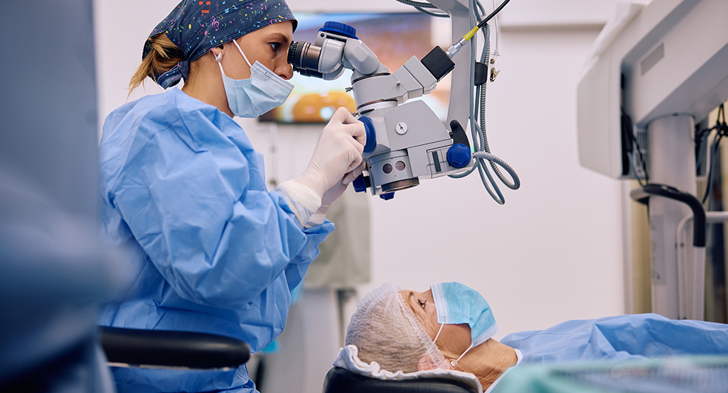
If you’re considering cataract surgery—or you’re a surgeon refining your technique—one topic that should be top of mind is endothelial cell loss. Why? Because the health of your corneal endothelium directly affects your vision after surgery. These delicate cells don’t regenerate, so once they’re damaged, they’re gone. And while cataract surgery is generally very safe, the way it’s done can have a huge impact on how well your eye heals—and how clear your vision stays in the long term.
In this article, we’ll break down the three big surgical factors that influence endothelial cell preservation: phacoemulsification power settings, incision size, and the type of viscoelastic material used. If you’re a patient, this will give you confidence to ask the right questions before your procedure. If you’re a clinician, you’ll find insights into how subtle changes in technique can significantly improve outcomes.
Why Does Endothelial Cell Loss Matter?
The corneal endothelium is a single layer of hexagonal cells lining the inner surface of your cornea. Their job? Pumping fluid out of the cornea to keep it clear. If too many of these cells are damaged during surgery, you can end up with corneal oedema and cloudy vision—a condition that can be temporary but sometimes becomes permanent.
Most patients experience a small degree of endothelial cell loss during cataract surgery—typically 4% to 10%. But certain factors can push that number much higher, especially in complicated cases or if older techniques are used. Understanding what influences this cell loss is the first step toward preventing it.
Phacoemulsification Power Settings: Getting the Balance Right
What Is Phacoemulsification?
Phacoemulsification (phaco) is the most common technique used to remove cataracts. It works by using ultrasonic energy to break up the cloudy lens inside your eye so it can be suctioned out. The trouble is, that same energy can also generate heat and turbulence—two things your corneal endothelium doesn’t like.
Lower Energy, Less Damage
Modern machines allow you to tailor the phaco settings in real-time. Using lower power settings, particularly with torsional or pulse modes, can drastically reduce the amount of ultrasound energy delivered into the eye. Less energy means less heat, less fluid movement, and a gentler environment for your endothelium.
Techniques That Help
Burst and pulse modes: Instead of continuous ultrasound, these deliver energy in short, controlled bursts—minimising turbulence.
Burst and pulse phaco modes are innovations designed to reduce the overall energy delivered into the eye during cataract surgery. Rather than relying on a constant stream of ultrasonic power, these modes allow for intermittent bursts that are carefully timed and measured.
This not only helps maintain better control over the lens emulsification process but also reduces the fluid turbulence and mechanical stress within the anterior chamber. In doing so, it lowers the chance of endothelial trauma—especially useful when operating near the corneal endothelium or in eyes with shallow anterior chambers.
The benefit of reduced turbulence is twofold: it safeguards the corneal endothelium and contributes to a more stable intraocular environment throughout the procedure. When fluid dynamics are better controlled, there’s less chance of chamber collapse, lens fragment bounce-back, or sudden pressure fluctuations—all of which can damage delicate endothelial cells. These settings also pair well with modern fluidics systems, which can precisely adjust flow and pressure in tandem with burst delivery, ensuring that the energy is used as efficiently and conservatively as possible.
Phaco chop vs divide-and-conquer: Phaco chop often uses less energy overall, making it more endothelial-friendly.
Phaco chop is a surgical technique where the lens nucleus is split into smaller fragments using a blunt instrument in conjunction with minimal phaco energy. Unlike the divide-and-conquer method—which requires sculpting deep grooves into the lens before cracking it—phaco chop allows for a quicker breakdown of the lens with less time spent delivering ultrasound energy.
This makes it a more energy-efficient approach, especially in dense cataracts where divide-and-conquer can demand prolonged phaco exposure. The reduced energy use significantly decreases the risk of collateral heat, fluid turbulence, and endothelial cell damage.
Additionally, the phaco chop method often involves less movement of the phaco tip, which further minimises agitation of the anterior chamber. Less manipulation inside the eye translates to fewer chances of accidental trauma to intraocular structures, including the corneal endothelium.
From a patient outcome perspective, the reduced energy and turbulence associated with phaco chop often result in faster visual recovery, lower postoperative inflammation, and more stable corneal clarity—especially important in patients with borderline endothelial counts.
Ozil torsional technology: This moves the tip in a side-to-side motion, rather than back-and-forth, reducing repulsion of lens fragments and cutting more efficiently.
Ozil torsional phaco technology introduces a side-to-side oscillating movement of the phaco tip, as opposed to the traditional longitudinal (back-and-forth) motion. This lateral action results in a smoother, more controlled emulsification of the lens material, reducing the tendency for lens fragments to be repelled away from the tip.
This reduction in repulsion means less energy is needed to keep fragments in position, which in turn decreases the overall turbulence and ultrasonic exposure within the anterior chamber.
For the corneal endothelium, this translates into a less hostile surgical environment. The smoother emulsification action helps maintain better chamber stability and reduces micro-shocks that could otherwise be transmitted toward the endothelium. Surgeons also find that torsional movement offers improved followability—the ability of the phaco tip to attract and retain lens material.
That combination of efficiency and control makes Ozil torsional technology particularly useful in preserving endothelial health, especially in complex cases involving dense nuclei or shallow anterior chambers.
Surgeon’s Role
Surgeon experience also plays a massive role. A skilled surgeon will modulate settings dynamically based on how the eye responds during surgery. The goal is always the same—remove the cataract efficiently while being as gentle as possible on the cornea.

Incision Size: Small Is Beautiful (But Not Always Simple)
Why Incision Size Matters
Making a small incision in the cornea is how we access the cataract. But the size and placement of that incision can influence how much fluid turbulence reaches the endothelium. Smaller incisions generally mean less disruption, less fluid leakage, and better maintenance of anterior chamber stability.
Micro-Incision Cataract Surgery (MICS)
MICS techniques typically use incisions smaller than 2.2 mm. Studies show that MICS is associated with significantly lower endothelial cell loss compared to standard incisions of 2.75 mm or larger. The smaller incision reduces outflow turbulence and maintains intraocular pressure more steadily during the procedure.
Trade-Offs and Technical Challenges
While smaller incisions sound ideal, they do come with trade-offs:
- Instrument flexibility: Working through a tight space can be more technically demanding.
- Wound integrity: It’s crucial the incision seals well to prevent postoperative complications.
Wound Architecture Matters Too
It’s not just the size—how the wound is constructed (bevelled vs straight, location, hydration technique) can make a difference. Poorly constructed wounds can cause unstable chambers, which in turn increase the likelihood of endothelial trauma.
Viscoelastic Agents: Cushioning the Endothelium
What Are They?
Viscoelastic materials are injected into the eye during surgery to protect the corneal endothelium and maintain space in the anterior chamber. Think of them as a physical buffer between surgical instruments and your precious endothelial cells.
Dispersive vs Cohesive: What’s the Difference?
- Dispersive viscoelastics (e.g., Viscoat) are better at coating and sticking to the corneal endothelium, making them excellent for protection.
- Cohesive viscoelastics (e.g., Healon) are better at maintaining space but don’t coat the endothelium as effectively.
Dual-Viscoelastic Techniques
Many surgeons now use a “soft-shell technique”—a combination of dispersive viscoelastic near the cornea and cohesive viscoelastic deeper in the chamber. This approach gives you the best of both worlds: optimal space maintenance and endothelial protection.
Timing and Technique
How and when you inject the viscoelastic matters too:
- It should be injected gently to avoid sudden pressure surges.
- Topping up during surgery helps maintain a consistent protective layer.
- Complete removal at the end is essential to avoid postoperative pressure spikes.
Other Factors That Influence Endothelial Health
Surgical Time
Longer surgeries generally lead to more cell loss. That’s why speed and efficiency—without rushing—are so important. Minimising time spent in the anterior chamber directly translates to better corneal outcomes.
Fluidics and Chamber Stability
Modern machines offer advanced fluidics systems that help maintain chamber stability. Sudden fluctuations in pressure can cause micro-trauma to the endothelium. The smoother the flow, the safer the procedure.
Pre-Existing Conditions
Some patients go into surgery with already compromised endothelium (e.g., Fuchs’ dystrophy). These patients require even more care—lower phaco settings, minimal surgical trauma, and sometimes even consideration of endothelial keratoplasty if damage is anticipated.
What the Research Shows
Multiple studies support the correlation between surgical technique and endothelial cell preservation:
Lower cumulative dissipated energy (CDE) is consistently linked to better endothelial outcomes.
Lower cumulative dissipated energy (CDE) means that less ultrasound energy is used during the phacoemulsification process. This is significant because excessive energy can lead to fluid turbulence, temperature rise, and mechanical stress within the anterior chamber—all of which put the corneal endothelium at risk.
When surgeons use lower CDE, they’re effectively reducing the physical toll on the intraocular environment, which directly translates to reduced trauma to the endothelial layer. This is especially relevant in cases with softer cataracts, where lower energy levels can still achieve sufficient lens fragmentation without overexertion of the system.
In real-world practice, optimising phaco settings to achieve the lowest effective CDE has become a hallmark of modern cataract surgery. Surgeons now use techniques like torsional phaco, pulse delivery, and phaco chop to lower energy use without compromising efficiency.
This refined approach not only shortens the duration of energy application but also limits the collateral impact on adjacent ocular tissues. Over time, a consistent focus on CDE optimisation contributes to improved corneal clarity, faster recovery, and better overall visual results for patients.
Incisions under 2.2 mm show reduced cell loss in comparative studies.
Reducing the size of the corneal incision may seem like a small detail, but the effect on endothelial cell preservation can be profound. Smaller incisions—typically under 2.2 mm—allow for better fluid control within the anterior chamber. This translates to less outflow turbulence, which otherwise might lead to microtrauma in the endothelial layer.
Furthermore, smaller wounds tend to seal more effectively, maintaining intraocular pressure and chamber depth more consistently during surgery. This mechanical stability reduces endothelial stress, especially during crucial steps like lens removal and IOL implantation.
However, the advantages of smaller incisions go beyond just fluidics. They also minimise postoperative inflammation and reduce the chance of surgically induced astigmatism, contributing to both functional and refractive success.
Importantly, adopting micro-incision cataract surgery (MICS) requires surgeon familiarity with modified tools and techniques, but once mastered, it becomes an effective strategy for maximising patient safety. In comparative clinical audits, eyes treated with incisions smaller than 2.2 mm consistently show lower rates of endothelial cell loss over short- and mid-term follow-up periods.
Dispersive viscoelastics have been shown to preserve 5–10% more endothelial cells in high-risk cases.
Dispersive viscoelastics, such as Viscoat, are particularly adept at coating and clinging to the inner corneal surface. This creates a protective cushion that guards against heat, shock waves, and mechanical trauma during phacoemulsification. In high-risk patients—like those with dense cataracts or compromised endothelial health—this extra layer of protection can make a significant difference.
The sticky, persistent nature of dispersive agents ensures that the endothelial layer remains shielded throughout the surgical procedure, even during high-flow irrigation or when ultrasonic energy is most intense.
The benefits of dispersive viscoelastics become especially evident when examining endothelial cell counts post-surgery. Comparative studies and surgeon-led case reviews have consistently shown a 5–10% improvement in endothelial preservation when dispersive viscoelastics are used over cohesive ones in vulnerable eyes.
This margin can be the difference between a smooth recovery and lingering corneal oedema. As such, many surgeons now adopt the soft-shell technique—combining dispersive and cohesive viscoelastics—to strike a balance between space maintenance and endothelial protection, optimising outcomes even in the most delicate of cases.
Final Thoughts: What You Can Do
If you’re a patient, you may not be adjusting phaco settings or choosing a viscoelastic—but you can absolutely ask the right questions. Is your surgeon using micro-incision techniques? Do they use advanced viscoelastics? How do they modulate energy delivery?
And if you’re a surgeon, these small tweaks in technique—combined with continual skill refinement—can dramatically improve outcomes for your patients. It’s not about reinventing the wheel. It’s about refining every touchpoint, every decision, every step.
Thinking of Cataract Surgery?
If you’re considering cataract surgery and would like to consult with an expert cataract surgeon, you can get in touch with us here at the London Cataract Centre. We’re happy to guide you through your options and help you find the safest, most effective treatment for your eyes.
References:
- Walkow, T., Anders, N., Klebe, S. and Wollensak, J., 2000. Endothelial cell loss after phacoemulsification: relation to preoperative and intraoperative parameters. Journal of Cataract and Refractive Surgery, 26(5), pp.727–732.
https://journals.lww.com/jcrs/Abstract/2000/05000/Endothelial_cell_loss_after_phacoemulsification_.17.aspx - Arshinoff, S.A. and Wong, E., 2003. Understanding and using the soft-shell technique. Journal of Cataract and Refractive Surgery, 29(5), pp.827–831.
- Bourne, R.R.A., Stevens, G.A., White, R.A., Smith, J.L., Flaxman, S.R., Price, H., Jonas, J.B., Keeffe, J., Leasher, J., Naidoo, K., Pesudovs, K. and Resnikoff, S., 2013. Causes of vision loss worldwide, 1990–2010: a systematic analysis. The Lancet Global Health, 1(6), pp.e339–e349.
https://www.thelancet.com/journals/langlo/article/PIIS2214-109X(13)70113-X/fulltext - Dick, H.B. and Augustin, A.J., 2001. Viscoelastics in ophthalmic surgery. Journal of Cataract and Refractive Surgery, 27(11), pp.1741–1749.
https://journals.lww.com/jcrs/Abstract/2001/11000/Viscoelastics_in_ophthalmic_surgery.12.aspx - Kohnen, T., Baumeister, M., Cichocki, M. and Kook, D., 2005. Impact of incision size on endothelial cell loss and surgically induced astigmatism after cataract surgery. Ophthalmologe, 102(10), pp.1039–1046.
https://link.springer.com/article/10.1007/s00347-005-1230-6