")

Cataract surgery is one of the most common and successful procedures performed worldwide. But when it comes to patients with Ehlers-Danlos Syndrome (EDS), things aren’t always so straightforward. If you or a loved one has been diagnosed with EDS and you’re facing cataract surgery, it’s entirely normal to have questions. The very nature of this connective tissue disorder means that certain risks are elevated—and that your surgical plan needs to be more nuanced.
In this article, we’re going to walk through everything you should know about how EDS impacts cataract surgery. From fragile ocular structures and bleeding tendencies to customised surgical strategies, we’ll look at how ophthalmologists work to ensure the safest possible outcomes.
What Is Ehlers-Danlos Syndrome and How Does It Affect the Eye?
Ehlers-Danlos Syndrome isn’t just one condition—it’s a group of hereditary disorders that affect the body’s connective tissues, which include skin, joints, and blood vessel walls. Because these tissues rely on collagen for strength and integrity, and collagen production is disrupted in EDS, various organs can become more fragile, including the eyes.
One of the key concerns in EDS, especially in its classical and vascular forms, is ocular fragility. The tissues in the eye—such as the sclera, cornea, and lens support structures—can be thinner or looser than normal. This can lead to complications like lens instability, corneal thinning, and a higher risk of retinal detachment.
For patients with EDS, even routine medical interventions can carry additional risks. Cataract surgery, although generally considered a low-risk procedure, must be approached with great care when EDS is involved. The surgeon has to take the patient’s unique anatomy into account to avoid complications and ensure a smooth recovery.
Zonular Weakness: When the Lens Loses Its Grip
One of the defining challenges in performing cataract surgery in someone with EDS is the issue of zonular weakness. Zonules are the tiny, thread-like fibres that hold your eye’s natural lens in place. In healthy eyes, they provide a stable scaffold, allowing the lens to sit perfectly centred inside the eye. In patients with EDS, these zonules can be loose, stretched, or even partially missing.
When the zonules are compromised, the lens can shift unpredictably. It might wobble during surgery or even be partially dislocated. This creates a surgical minefield for the ophthalmologist. Standard techniques that rely on stable lens positioning may not work well, and there’s a greater risk that the lens capsule could rupture.
To navigate this, surgeons often turn to devices like capsular tension rings (CTRs). These are small circular implants inserted into the lens capsule to help redistribute tension and support the structure. In some cases, they may even use segmental devices that anchor the capsule to the sclera for extra support. These modifications allow for a safer procedure despite the fragility of the ocular infrastructure.
Increased Risk of Bleeding: When Fragile Vessels Come Into Play
Another major issue in EDS is increased bleeding risk. While not all types of EDS affect blood vessels, the vascular subtype in particular is notorious for making vessel walls prone to rupture. Even in other types, bruising and bleeding can occur more readily than usual, particularly if the patient has associated platelet dysfunction or fragile capillaries.
During cataract surgery, bleeding is typically minimal. But in EDS, even small incisions can result in more extensive haemorrhaging, especially if care isn’t taken. Subconjunctival haemorrhages (bleeding under the white of the eye) may look alarming, even if they aren’t dangerous. However, deeper bleeding within the eye—such as from the iris or ciliary body—can increase intraoperative difficulty and postoperative inflammation.
Surgeons can mitigate this by choosing anaesthetic techniques that reduce blood pressure spikes and by using medications like tranexamic acid (where safe and indicated) to reduce bleeding risk. Careful preoperative blood work and discussions with a haematologist may also be part of the planning process.
Wound Healing and Dehiscence: The Threat of Sutures Giving Way
Poor wound healing is another hallmark concern in Ehlers-Danlos Syndrome. Connective tissue that doesn’t repair itself properly can lead to wound dehiscence, where surgical incisions begin to reopen after closure. In a sensitive area like the eye, that can be catastrophic.
The tiny incisions made during modern cataract surgery are usually self-sealing. But in EDS patients, these wounds might not stay closed on their own. Surgeons may opt to place sutures even in surgeries that would normally not require them. More importantly, these sutures need to be placed very carefully so as not to cause further tearing of fragile scleral or corneal tissue.
Using finer, slower-absorbing suture materials and meticulous surgical technique is key. In the postoperative period, these patients also need to be closely monitored. Any signs of wound leakage, pain, or inflammation must be taken seriously and addressed promptly to prevent vision-threatening complications.
Surgical Planning: Preoperative Steps That Make a Big Difference
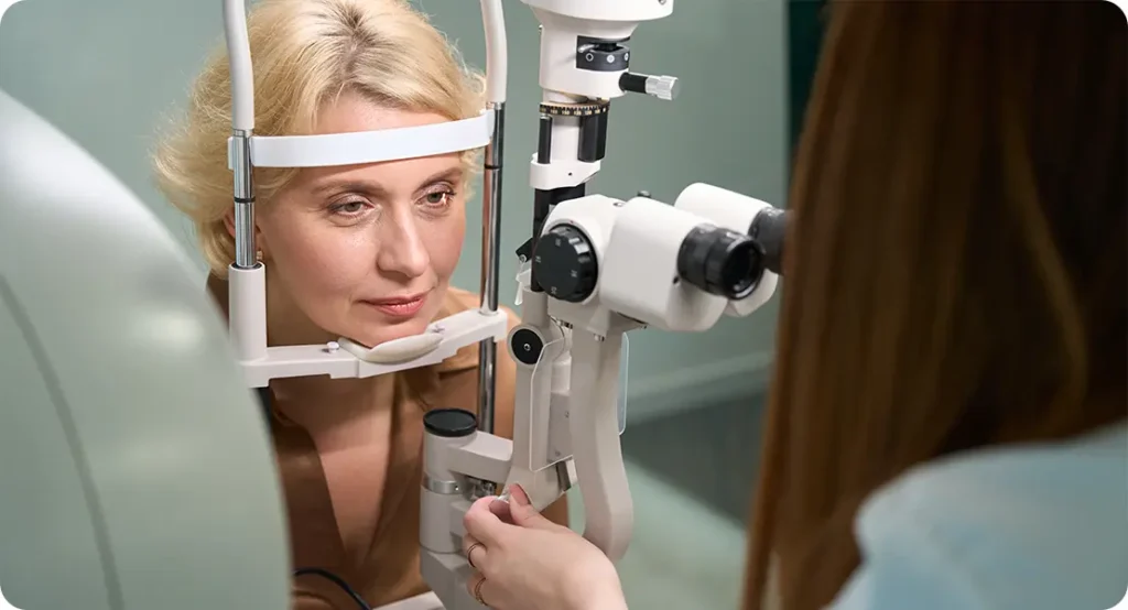
Before any surgery begins, your eye surgeon will carry out a detailed preoperative assessment. In patients with EDS, this step becomes even more critical. Expect additional imaging, such as anterior segment OCT (optical coherence tomography), to evaluate the integrity of your eye’s structures. The surgeon will also want to know your full medical history, including any prior ocular surgeries, your EDS subtype, and whether you have vascular involvement.
The surgical plan may involve changes in technique, like choosing a slightly larger or more reinforced incision, selecting different types of intraocular lenses (IOLs), and pre-positioning instruments to manage potential lens instability. Anaesthetic planning also becomes important—some surgeons will collaborate with anaesthetists familiar with connective tissue disorders to ensure a smooth and stable procedure.
Patients may also be asked to temporarily stop medications that affect bleeding (with approval from their GP or specialist), or to begin prophylactic antibiotic or anti-inflammatory eye drops a few days before surgery to reduce infection and inflammation risk.
Intraoperative Adjustments: Flexibility in the Operating Room
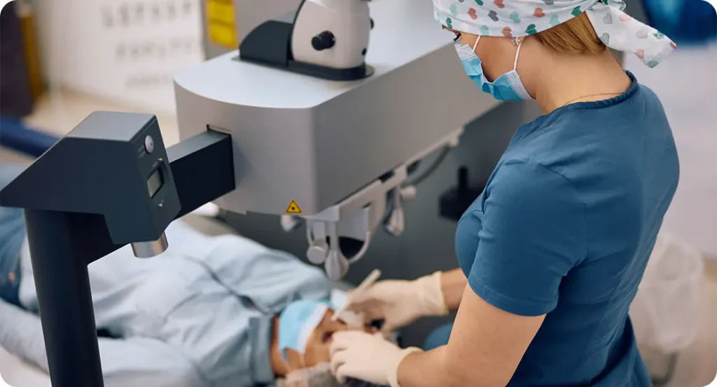
No matter how well the surgery is planned, there’s always the chance that something unexpected will happen during the operation—particularly when EDS is involved. That’s why surgeons need to be prepared to make intraoperative adjustments on the fly.
This might mean switching from a standard IOL to one that can be scleral-sutured in place if the lens capsule proves unstable. Or it could involve using viscoelastic substances more generously to protect delicate corneal tissues. Surgeons might need to make smaller, incremental incisions or take longer to perform each step to avoid stressing fragile structures.
These surgeries tend to take a little longer, and precision matters even more than usual. However, with careful attention and the right tools at hand, the outcomes can still be excellent.
Postoperative Recovery: A Time for Close Observation
Once the surgery is complete, the next critical stage begins—recovery. In EDS patients, this isn’t just about checking that the lens is sitting well and vision is improving. It’s about watching closely for signs of wound leakage, inflammation, infection, or unexpected lens movement.
You’ll likely be asked to come in for more frequent postoperative check-ups compared to standard cataract patients. These might happen on day one, week one, and again at regular intervals through the first few months. Your doctor will be looking for even subtle signs that the eye isn’t healing as expected.
You may be advised to use anti-inflammatory drops for a longer period of time than usual. Additionally, physical activity may need to be limited during the healing period to avoid any accidental eye trauma or straining. Wearing a protective shield at night might also be recommended for longer.
Long-Term Visual Outcomes: What Can You Expect?
The good news is that, despite the added challenges, many patients with EDS who undergo cataract surgery do achieve excellent visual results. The keys lie in the preoperative planning, tailored surgical technique, and attentive postoperative care.
However, some patients may experience slightly longer visual recovery times or require additional procedures, especially if complications such as lens decentration or capsular opacification occur. In rare cases, further interventions such as YAG laser capsulotomy or even IOL repositioning may be necessary.
The bottom line? With the right surgical team and approach, there’s every reason to be optimistic about the outcome.
Psychological Preparation: Managing Expectations and Anxiety
Facing surgery with a complex medical condition like EDS can understandably be stressful. Some patients worry about the “what-ifs” more than the procedure itself. It’s completely natural to feel this way.
Open communication with your eye surgeon is essential. Make sure you understand what adjustments will be made to the standard procedure, and what steps are in place to keep you safe. Don’t hesitate to ask about the surgeon’s experience with connective tissue disorders and whether other specialists (such as anaesthetists or haematologists) will be involved.
Sometimes, arranging a preoperative consultation with a counsellor or patient advocate can also be helpful. Having someone to talk to about your concerns may ease the emotional burden and give you more confidence going into surgery.
Collaborative Care: The Role of Multidisciplinary Teams
Managing cataract surgery in EDS patients isn’t just a job for the ophthalmologist. In many cases, it becomes a team effort. Your GP, geneticist, rheumatologist, haematologist, and even a cardiologist may be consulted depending on your medical history.
A multidisciplinary approach ensures that every aspect of your health is considered—from your cardiovascular stability to your clotting profile to your ability to heal from surgical wounds. This team-based care is especially important for patients with vascular EDS, where complications can be life-threatening if not anticipated and managed correctly.
The goal is to make surgery not only safer but also more successful in the long term.
Lifestyle Tips After Surgery for EDS Patients
Even once the eye has healed and your vision has improved, it’s wise to make a few lifestyle adjustments to protect your results. If you wear glasses, make sure the frames are comfortable and don’t exert undue pressure on your temples or nose bridge—areas that might bruise more easily.
Stay hydrated, eat a nutrient-rich diet that supports collagen and tissue repair, and avoid any eye-rubbing habits. Regular eye exams should remain a part of your healthcare routine, not just for vision monitoring but to check on the positioning and clarity of your intraocular lens.
Some patients also benefit from physical therapy or low-impact exercises tailored to EDS, which can help maintain overall health without putting too much strain on joints or connective tissues.
FAQs
- Is cataract surgery safe for patients with Ehlers-Danlos Syndrome?
Yes, cataract surgery can be safely performed in patients with Ehlers-Danlos Syndrome, but it requires a highly tailored approach. The unique risks related to tissue fragility, loose zonules, and bleeding tendencies mean that standard techniques are often modified to minimise complications. Surgeons with experience in connective tissue disorders will typically make strategic adjustments before, during, and after the procedure to improve safety and outcomes. - What are the biggest risks during cataract surgery for someone with EDS?
The main risks include zonular weakness leading to lens instability, fragile tissues causing tears or dehiscence, and an increased tendency for bleeding. These risks can complicate the procedure and prolong recovery if not properly managed. However, with appropriate preoperative imaging, surgical planning, and the use of specialised equipment like capsular tension rings, most of these risks can be significantly reduced. - Will I need stitches after cataract surgery if I have EDS?
While many cataract surgeries do not require sutures thanks to self-sealing incisions, patients with EDS often have weaker tissue integrity, which can make these wounds less reliable. As a precaution, surgeons may choose to place fine, absorbable stitches to secure the incision and prevent it from reopening, especially in cases where the sclera or cornea is particularly thin or delicate. - How does zonular weakness affect the surgery?
Zonular fibres are crucial for keeping the lens in place, and in EDS they can be stretched, loose, or absent. This makes lens removal and replacement more challenging, as the capsule may not be stable enough on its own. To counter this, surgeons may use capsular tension rings or other stabilising devices to support the capsule during the procedure and ensure correct intraocular lens positioning. - Can EDS cause complications with the intraocular lens (IOL)?
Yes, if the lens capsule is unstable or weak due to loose zonules, the intraocular lens might shift or decenter over time. Surgeons address this by selecting IOLs that are more stable in compromised capsules or by fixing them to the sclera when necessary. Postoperative monitoring is also more frequent to ensure the lens stays properly positioned as the eye heals. - What precautions are taken to prevent excessive bleeding during surgery?
Surgeons will often assess your clotting profile in advance and may coordinate with a haematologist if you’re at higher bleeding risk, especially in vascular EDS. During surgery, they use gentle tissue handling, meticulous technique, and may employ vasoconstrictive agents or other medications to control bleeding. Care is also taken during anaesthesia to avoid sudden blood pressure spikes that could stress fragile vessels. - How long is the recovery period for EDS patients after cataract surgery?
The recovery time can be longer than usual due to slower wound healing and the potential for inflammation or complications. Patients are typically seen more frequently in the first few weeks and may be kept on anti-inflammatory eye drops for an extended period. Full visual recovery may take several weeks or even months, but careful monitoring ensures any issues are caught early. - Will I need a general anaesthetic instead of local?
Most cataract surgeries, even in EDS patients, are done under local anaesthesia, which reduces systemic risk and allows for faster recovery. However, if the patient has complex medical needs or significant anxiety, general anaesthesia might be considered. Anaesthetic plans are carefully tailored in collaboration with an experienced anaesthetist to ensure cardiovascular and airway safety, particularly in those with vascular fragility. - Should I stop any medications before the surgery?
Some medications, particularly those that affect bleeding (like aspirin or blood thinners), may need to be paused or adjusted prior to surgery. However, this should always be done under medical supervision, ideally in consultation with your GP or specialist. Your ophthalmologist will discuss any required changes during your preoperative assessment to balance the risks of bleeding and clotting. - How do I choose a surgeon if I have Ehlers-Danlos Syndrome?
Choosing a surgeon who has experience with connective tissue disorders is key. Look for an ophthalmologist who understands the specific risks associated with EDS and is willing to personalise the surgical approach accordingly. It’s also important that the surgical centre has access to advanced imaging and intraoperative tools to manage potential challenges effectively and safely.
Final Thoughts
Cataract surgery in patients with Ehlers-Danlos Syndrome requires thoughtful, customised planning—but that doesn’t mean it has to be risky or overwhelming. By working with an experienced surgeon and following a carefully structured pre- and post-operative plan, most people with EDS can look forward to clear vision and smooth healing.
At London Cataract Centre, we understand the intricacies of treating patients with complex connective tissue disorders like EDS. Our team takes a meticulous, individualised approach to every case, prioritising both safety and long-term outcomes. If you or someone you know is preparing for cataract surgery and has EDS, don’t hesitate to get in touch with a specialist team who understands the full picture.
References
1. Malfait, F., Francomano, C.A., Byers, P.H., Belmont, J., Berglund, B., Black, J., Bloom, L., Bowen, J.M., Brady, A.F., Burrows, N.P. and Castori, M., 2017. The 2017 international classification of the Ehlers–Danlos syndromes. American Journal of Medical Genetics Part C: Seminars in Medical Genetics, 175(1), pp.8–26.
Available at: https://onlinelibrary.wiley.com/doi/full/10.1002/ajmg.c.31552 [Accessed 30 Jun. 2025].
2. Atik, T., 2022. Ehlers-Danlos syndrome: current perspectives. The Application of Clinical Genetics, 15, pp.21–37.
Available at: https://www.ncbi.nlm.nih.gov/pmc/articles/PMC8904282/ [Accessed 30 Jun. 2025].
3. Germain, D.P., 2007. Ehlers-Danlos syndrome type IV. Orphanet Journal of Rare Diseases, 2(32), pp.1–9.
Available at: https://ojrd.biomedcentral.com/articles/10.1186/1750-1172-2-32 [Accessed 30 Jun. 2025].
4. Grzybowski, A., Kanclerz, P., Muzyka-Woźniak, M. and Adamiec-Mroczek, J., 2020. Modern cataract surgery: safety, effectiveness and new trends. Journal of Clinical Medicine, 9(11), p.3474.
Available at: https://www.mdpi.com/2077-0383/9/11/3474 [Accessed 30 Jun. 2025].