")

Cataracts are a common eye condition characterised by the clouding of the eye’s natural lens, which leads to blurred or distorted vision. Among the different types of cataracts, cortical cataracts are distinctive due to their location and appearance. They develop in the lens cortex—the outer layer of the lens—and typically begin as white, wedge-shaped opacities that point inward toward the centre of the eye. This article explores the features, causes, symptoms, diagnostic approaches, and treatment options related to cortical cataracts.
What Are Cortical Cataracts?
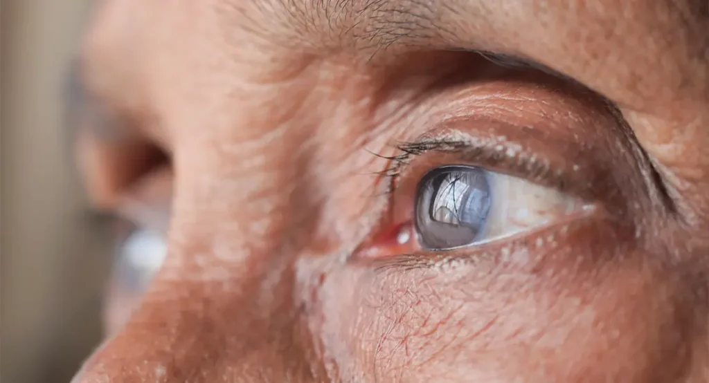
Cortical cataracts develop in the cortex of the lens—the outermost layer made up of elongated, fibre-like cells arranged in a radial pattern. These cataracts typically begin as small, white or greyish opacities located around the outer edge of the lens. Over time, they spread inwards toward the centre in a wedge-shaped or spoke-like configuration. This pattern is quite distinctive and can be observed during a slit-lamp examination. Because the cortical region is responsible for allowing light to pass through to the central part of the retina, any disruption in this area can eventually distort how light is focused in the eye.
In contrast to nuclear cataracts, which start in the central core of the lens and tend to progress slowly by yellowing and hardening, cortical cataracts often present more erratically. Their peripheral location means that in the initial stages, central vision may remain relatively unaffected. However, the irregularity of the opacities can cause light to scatter unpredictably within the eye, leading to early signs of visual discomfort even when overall visual acuity remains normal. This scattering effect is not uniform and may vary depending on lighting conditions, making the symptoms of cortical cataracts particularly frustrating for patients.
As the cataract continues to progress, the radial spokes grow larger and more numerous, eventually converging towards the centre of the lens. When this happens, light entering the eye becomes increasingly disrupted, affecting both near and distance vision. Patients may start to notice a loss of sharpness or clarity, especially when trying to read or recognise faces. Peripheral vision might also become compromised, which can impact tasks requiring wide visual awareness, such as driving. This gradual reduction in visual function tends to occur unevenly, often leading to difficulty adapting between different lighting environments.
One of the most troubling symptoms associated with cortical cataracts is increased sensitivity to glare. Because the lens is no longer able to filter and focus light evenly, bright lights—particularly headlights at night or sunlight reflecting off surfaces—can cause visual discomfort, haloes, or even temporary blinding. This symptom can significantly reduce quality of life and limit a person’s ability to perform everyday activities safely. The erratic light scattering can also make it harder to distinguish contrast, especially in dim conditions, further highlighting the unique visual challenges posed by this type of cataract.
Causes and Risk Factors
Cortical cataracts are usually age-related, most commonly developing in people over the age of 60. However, several other contributing factors may accelerate their onset or worsen their progression:
Ageing
As the body ages, natural biochemical changes occur in various tissues, including the lens of the eye. Over time, the proteins within the lens begin to denature and clump together, disrupting its clarity. These changes are gradual and often go unnoticed at first but can eventually lead to visible opacities, particularly in the outer cortical region.
Oxidative stress plays a central role in age-related lens damage. The lens is constantly exposed to light and oxygen, which, while essential for vision, also contribute to the generation of free radicals. These unstable molecules can damage lens fibres, reducing their transparency and elasticity, especially when the body’s natural antioxidant defences begin to decline with age.
Furthermore, the lens loses its ability to regulate hydration and nutrient exchange efficiently as it gets older. The disruption of fluid balance within the lens fibres can cause them to swell and break down. This breakdown contributes to the formation of wedge-shaped cortical opacities, which interfere with normal light transmission through the lens.
Diabetes
Diabetes is a significant risk factor for cortical cataracts, primarily due to the effect of chronic high blood sugar on lens metabolism. Elevated glucose levels lead to an accumulation of sorbitol within the lens via the polyol pathway. This sugar alcohol draws water into the lens fibres, causing them to swell and become cloudy.
The metabolic changes in diabetic patients also result in oxidative stress within the lens. Excess glucose alters the redox balance, reducing the availability of antioxidants like glutathione, which protect the lens proteins from damage. This imbalance makes the lens more vulnerable to opacification, especially in the cortical zone.
Moreover, diabetes can affect the microcirculation of the eye, impairing the delivery of nutrients to the lens. When the lens doesn’t receive adequate nourishment, its natural repair mechanisms falter, increasing the risk of protein aggregation and structural irregularities. This damage manifests as cortical clouding that often progresses more rapidly than in non-diabetic individuals.
Excessive Sun Exposure
Prolonged exposure to ultraviolet light, particularly UV-B rays, is known to accelerate lens damage. UV radiation can alter the molecular structure of the lens proteins, causing them to unfold and clump together. This structural disruption typically begins in the lens cortex, where the light enters, and can lead to characteristic spoke-like opacities.
The lens does have some natural defences against UV damage, including the presence of protective chromophores that absorb UV radiation. However, with repeated exposure over years, these defences are overwhelmed. The resulting photo-oxidative stress triggers inflammatory and degenerative changes within the lens fibres.
People who spend significant time outdoors without adequate eye protection are at greater risk. Wearing sunglasses with proper UV filtration can significantly reduce the risk, but once the damage has occurred, it is irreversible. Cortical cataracts related to sun exposure often present earlier than age-related types and may progress more aggressively.
Smoking
Smoking introduces a cocktail of harmful chemicals into the bloodstream, many of which can reach the delicate tissues of the eye. The lens is particularly susceptible to the oxidative damage caused by tobacco smoke, which depletes antioxidants and encourages the formation of protein clumps within the lens cortex.
Nicotine and other compounds in cigarettes promote chronic inflammation and vascular constriction. These effects can limit the delivery of oxygen and nutrients to the lens, impairing its ability to maintain transparency. Over time, this can lead to the development of cortical cataracts, even in relatively young individuals.

Studies have also shown that smokers tend to have lower levels of key antioxidants in the eye, including vitamin C and glutathione. Without these protective substances, the lens is more prone to oxidative damage. The impact of smoking is cumulative, meaning the longer one smokes, the higher the risk of cataract formation—particularly in the peripheral regions of the lens.
Hypertension
High blood pressure affects more than just the heart and arteries—it can also impair ocular health. The lens relies on a steady supply of nutrients delivered through the aqueous humour, which is influenced by the vascular health of nearby structures. Elevated blood pressure can disrupt this balance, leading to subtle but damaging changes in lens metabolism.
Hypertension can contribute to oxidative stress by damaging small blood vessels throughout the body, including those that support the eye. When these vessels become less efficient, waste removal from the lens is compromised. The accumulation of metabolic byproducts can increase the likelihood of lens opacities, particularly in the cortical area.
In addition, hypertension is often associated with other risk factors such as obesity and diabetes, which together can create a compounded risk for cataract development. Managing blood pressure through lifestyle changes and medication may therefore have a protective effect on the eyes, delaying the onset of cortical cataracts.
Genetics
A family history of cataracts suggests that genetic factors play a role in determining an individual’s risk. Certain inherited mutations can affect how the lens develops, repairs itself, or responds to environmental stressors. These mutations may impair the structure or function of crystallin proteins, which are essential for maintaining lens clarity.
Some people inherit a reduced capacity to neutralise oxidative stress due to variations in genes that code for antioxidant enzymes. This makes their lenses more susceptible to damage from UV light, free radicals, and other environmental insults. The result is an increased likelihood of developing cortical opacities, sometimes at an earlier age than is typical.
While genetic predisposition cannot be changed, it can be accounted for through regular monitoring and preventive care. Individuals with a strong family history of cataracts may benefit from earlier and more frequent eye examinations, combined with proactive lifestyle adjustments to minimise avoidable risks.
Trauma or Eye Injury
Physical injury to the eye can disturb the precise organisation of lens fibres, leading to the formation of cataracts. A blunt or penetrating trauma may cause sudden structural damage that interrupts the clarity of the lens cortex. In some cases, cortical cataracts can form shortly after the injury; in others, they may develop slowly over time.
The trauma often induces a localised inflammatory response, which can disrupt the biochemical environment of the lens. This inflammation, combined with possible mechanical disruption, contributes to the opacification of lens tissue. Cortical opacities resulting from trauma may be irregular in shape and location, often differing from the typical spoke-like pattern seen in age-related cases.
Even seemingly minor injuries can have long-term consequences. For instance, a sports-related blow or accidental poke to the eye might not cause immediate symptoms but could initiate subtle changes that culminate in cataract formation years later. Protective eyewear during high-risk activities is essential to minimise such risks.
Use of Corticosteroids
Corticosteroids, while medically useful for a variety of conditions, are known to have ocular side effects when used long-term. These medications may interfere with the normal maintenance and repair of lens fibres by altering protein synthesis and promoting oxidative damage within the eye.
Topical, systemic, or inhaled corticosteroids have all been implicated in cataract development, although the risk appears to be dose- and duration-dependent. Cortical cataracts caused by steroid use often present bilaterally and may progress silently before causing noticeable symptoms.
The mechanism is not fully understood, but one theory suggests that corticosteroids disrupt the balance of electrolytes and water within the lens, affecting its transparency. Patients who require chronic corticosteroid therapy should be closely monitored by an eye care professional, with routine examinations to detect early signs of lens changes.
Symptoms of Cortical Cataracts
Cortical cataracts may initially go unnoticed, especially if the central vision remains unaffected. As the condition progresses, patients may begin to notice the following symptoms:
- Blurred or hazy vision, particularly in bright light: In the early stages, blurred vision caused by cortical cataracts can be intermittent and mild, often mistaken for the need for new glasses or tired eyes. The haze typically stems from the light scattering caused by wedge-shaped opacities around the lens, which disrupt the smooth passage of light onto the retina. As more spokes form and converge towards the centre, this blurring can worsen, affecting visual clarity even in well-lit conditions. This symptom often becomes more pronounced in bright environments, such as outdoors during daylight or in brightly lit rooms. Paradoxically, instead of seeing better with more light, individuals with cortical cataracts may find their vision becomes more washed out or obscured. The increased brightness heightens the scattering effect of the opacities, which can cause discomfort and reduce visual detail.
- Glare sensitivity, especially when driving at night: One of the more troubling symptoms for patients is increased glare sensitivity, particularly during night-time driving. Headlights from oncoming traffic may appear overly bright, distorted, or smeared, creating visual confusion and discomfort. This occurs because the cortical opacities scatter light from multiple sources, leading to overlapping images and difficulty focusing. As glare sensitivity increases, many people begin to avoid driving at night altogether due to safety concerns. Even familiar roads can feel treacherous when lights cause temporary blindness or disorientation. This can significantly impact independence, especially for older adults, limiting their ability to socialise or carry out evening tasks confidently.
- Halos around lights: Patients with cortical cataracts often report seeing halos or rings around lights, especially in dim environments. These halos are usually the result of irregular diffraction and refraction of light by the disrupted lens fibres. Streetlamps, car headlights, and even indoor lighting can appear to have bright rings or smudges surrounding them, which can interfere with visual focus. These halos can be particularly disorienting at night or in low-contrast settings, when the eye relies heavily on clear light cues to navigate the environment. Unlike the mild glare experienced by some people without cataracts, the halos caused by cortical opacities are more persistent and often accompanied by visual discomfort, making daily activities more challenging.
- Difficulty with contrast, making it harder to distinguish between shades of similar colours: Cortical cataracts can reduce the eye’s ability to perceive contrast, making it difficult to tell apart objects or surfaces with similar tones. For example, cream-coloured writing on a white background or grey pavement against a similarly coloured curb may appear blended. This loss of contrast sensitivity results from the lens’s inability to focus light cleanly, leading to a ‘flattening’ of visual information. This impairment can become problematic when navigating unfamiliar environments, reading low-contrast text, or performing tasks that require fine visual discrimination, such as choosing matching clothes or reading instruments. Over time, this diminished contrast sensitivity can contribute to missteps or accidents, especially in poorly lit areas.
- Double vision in one eye, which is a result of irregular light refraction through the cloudy lens: Monocular double vision—seeing two overlapping images in one eye—is a less common but distinctive symptom of cortical cataracts. It typically arises when light is refracted inconsistently by the spoke-like opacities in the lens cortex. Unlike binocular double vision, which usually stems from eye muscle misalignment, this type is caused by the distorted transmission of light within a single lens. This symptom can be especially confusing and may not resolve even when one eye is closed. Reading, recognising faces, or focusing on fine detail may become difficult. Patients might describe seeing a shadow image alongside the real one, making vision feel unstable or wobbly. It often persists until the cataract is treated surgically, at which point clarity usually returns rapidly.
Diagnosis
The diagnosis of cortical cataracts involves a thorough eye examination by an ophthalmologist or optometrist. Key components of the assessment include:

- Slit-lamp examination: The slit-lamp examination is a fundamental tool in cataract diagnosis. It uses a high-intensity light and microscope to illuminate and magnify the structures of the eye, allowing the clinician to see the lens in cross-section. This technique reveals the presence, size, and distribution of the wedge-shaped cortical opacities, which often appear as white streaks or spokes radiating toward the centre of the lens. This method also enables the practitioner to differentiate cortical cataracts from other types, such as nuclear or posterior subcapsular cataracts, by observing their specific pattern and location. The examination is painless and usually takes just a few minutes, but its detail and accuracy are essential for deciding the appropriate course of management.
- Visual acuity testing: Visual acuity testing measures how clearly a person can see letters or symbols at a standard distance. It helps quantify the extent to which the cataract is impairing sight, particularly in terms of sharpness and clarity. The results are usually expressed as a fraction (e.g., 6/12), which provides a baseline for monitoring disease progression. In cases of cortical cataracts, patients might show relatively good vision under normal lighting but significantly worse performance in high-glare or low-contrast environments. This discrepancy can alert the clinician to the presence of cortical involvement, even when the central lens appears mostly clear.
- Dilated eye examination: During a dilated eye exam, special drops are used to widen the pupil, giving the clinician a broader view of the internal eye structures. This is particularly important for assessing the full extent of the cataract, as cortical opacities often originate in the periphery and may be missed with an undilated pupil. In addition to evaluating the lens, dilation allows the examiner to inspect the retina and optic nerve, ensuring that no other underlying conditions—such as macular degeneration or glaucoma—are contributing to the patient’s symptoms. This comprehensive approach helps guide accurate diagnosis and treatment planning.
- Glare testing: Glare testing assesses how much bright light interferes with a patient’s vision. It simulates real-world lighting conditions, such as oncoming headlights or midday sun, and measures how well the eyes can perform under these circumstances. This is particularly useful for detecting functional vision loss that might not be captured through standard acuity tests. Cortical cataracts often scatter light in unpredictable ways, making this test highly relevant. A patient may have decent vision on a letter chart but struggle in bright environments. Glare testing helps validate these experiences and can be an important factor in deciding when surgical intervention is warranted.
Management and Treatment
In the early stages, when symptoms are mild, non-surgical strategies may be sufficient. These may include:

- Updated prescription lenses: As vision starts to decline due to cortical cataracts, updating glasses or contact lens prescriptions can provide temporary relief. Minor adjustments in lens strength or refraction can help refocus light more accurately through the partially clouded lens, improving clarity for tasks like reading or computer work. However, while new lenses may help sharpen vision, they do not slow the progression of the cataract itself. Regular eye checks remain important, as prescriptions may need to be changed more frequently than before. This strategy is most effective in the early phase, when cataracts have not yet reached the visual axis.
- Use of anti-glare sunglasses: Anti-glare or polarised sunglasses can be a simple yet highly effective tool for managing glare sensitivity, one of the hallmark symptoms of cortical cataracts. These lenses are designed to reduce the amount of scattered light entering the eye, making outdoor activities more comfortable and safer. Wearing them during the day—especially when driving or walking in bright sunlight—can significantly improve contrast and reduce visual strain. Although they don’t treat the cataract, they help compensate for one of its most troublesome effects and enhance day-to-day functionality.
- Brighter lighting at home or work: Increased ambient lighting, particularly with high-contrast, warm-coloured light sources, can counteract some of the visual dullness caused by cortical cataracts. It becomes easier to see fine details, such as small print or stitches in fabric, when the environment is well illuminated. Adjustments like adding reading lamps, using daylight-spectrum bulbs, or installing under-cabinet lighting in kitchens can make a noticeable difference. These measures are often recommended alongside other conservative treatments to maintain independence in daily tasks.
- Avoidance of night driving: Driving at night is one of the first activities many patients with cortical cataracts begin to struggle with. Headlights, streetlights, and reflective signs can create intense glare and haloes, impairing reaction times and depth perception. As a result, it may be safer to avoid driving after dark, especially on unfamiliar roads or during inclement weather. By recognising this limitation early, patients can make practical adjustments—such as arranging rides or planning travel during daylight hours—before the situation becomes dangerous. It is also a useful indicator for eye care professionals to assess when vision loss is beginning to impact quality of life.
Cataract Surgery
Cataract surgery is a highly effective and commonly performed procedure. The clouded lens is removed and replaced with a clear artificial intraocular lens (IOL). The operation is typically done under local anaesthesia and takes less than 30 minutes in most cases.

Cortical cataracts can pose unique challenges during surgery due to the irregular lens opacities, but with modern techniques such as phacoemulsification and advanced IOL options, outcomes are generally excellent.
Prognosis and Prevention
With prompt diagnosis and appropriate treatment, the prognosis for patients with cortical cataracts is very good. Visual outcomes following surgery are often dramatic, with many individuals regaining near-normal vision.
While not all cortical cataracts can be prevented, certain lifestyle adjustments may help delay their onset:
- Wearing UV-protective sunglasses outdoors
- Maintaining good control of systemic conditions, especially diabetes and hypertension
- Avoiding smoking
- Eating a diet rich in antioxidants, such as leafy greens, berries, and nuts
- Having regular eye examinations, particularly after the age of 50
Conclusion
Cortical cataracts represent a common yet manageable form of age-related lens opacity. Although they may not cause noticeable symptoms in the early stages, their progression can lead to significant visual impairment. Fortunately, both non-surgical and surgical treatment options are available, with excellent outcomes when appropriately managed. Maintaining regular eye check-ups and adopting healthy lifestyle choices can go a long way in preserving clear vision well into later life.
If you are concerned about cortical cataracts or experiencing any changes in your vision, you can contact us at the London Cataract Centre to arrange a consultation with one of our cataract surgeons.