")

If you have been diagnosed with angle-closure glaucoma, it is natural to wonder whether cataract surgery could help manage your condition. Many people assume cataract surgery is only carried out to improve blurred vision. In reality, it can also play an important role in controlling eye pressure. This is particularly relevant when glaucoma is driven by eye anatomy rather than fluid resistance alone.
Angle-closure glaucoma behaves differently from other forms of glaucoma. The drainage angle becomes narrow or blocked, preventing fluid from leaving the eye efficiently. As a result, eye pressure can rise and place the optic nerve at risk. Because this mechanism is structural, treatment often focuses on creating more space inside the eye.
The natural lens thickens as it ages and gradually pushes the iris forwards. This further narrows the drainage angle and increases the likelihood of pressure build-up. Removing the cataract lens creates more room in the front of the eye. This can help reopen the angle and improve fluid drainage.
For many patients, cataract surgery therefore offers both visual and pressure-related benefits. It may reduce the risk of future pressure spikes and lessen reliance on glaucoma medication. While it is not suitable for everyone, careful assessment helps determine whether this approach is appropriate. Understanding this connection allows you to make informed decisions about your long-term eye care.
Understanding Angle-Closure Glaucoma
Angle-closure glaucoma occurs when the angle between the iris and cornea becomes too narrow. This drainage angle is vital for allowing fluid to leave the eye normally. When it becomes blocked, eye pressure can rise rapidly or increase slowly over time. Both patterns can damage the optic nerve if not treated promptly. Early recognition and careful management are therefore essential.
Unlike open-angle glaucoma, angle-closure glaucoma is strongly influenced by the physical structure of the eye. Certain anatomical features make some people more vulnerable. These risks often increase with age as natural changes occur within the eye. Structural factors play a central role in how and when the condition develops.
As the natural lens thickens over time, it can push the iris forward. This reduces the space in the drainage angle and restricts fluid outflow. Pressure may then rise without warning or progress quietly. Regular eye examinations help detect these changes before permanent damage occurs.
Key Risk Factors for Angle-Closure Glaucoma
- Naturally shallow front chamber of the eye
- Smaller overall eye size
- Thickening of the natural lens with age
- Narrow drainage angles on examination
- Family history of angle-closure glaucoma
Acute and Chronic Angle-Closure Glaucoma
Angle-closure glaucoma can present suddenly or develop gradually. Acute angle closure causes severe symptoms such as eye pain, blurred vision, headaches, and nausea. This is a medical emergency that requires immediate treatment. Without urgent care, permanent vision loss can occur quickly.
Chronic angle-closure glaucoma develops more quietly. Eye pressure rises slowly, and symptoms may be mild or completely absent. Damage can progress before it is noticed, which is why regular eye examinations are extremely important. Early detection allows treatment before significant vision loss occurs.
Key Differences Between Acute and Chronic Angle-Closure Glaucoma
- Acute angle closure develops suddenly and causes severe symptoms
- Chronic angle closure progresses slowly with few or no early symptoms
- Acute attacks require emergency treatment
- Chronic disease is often detected during routine eye examinations
- Both forms can lead to permanent vision loss if untreated
Why Cataracts Worsen Angle Closure
Cataracts form as the natural lens becomes thicker and less transparent over time. As the lens enlarges, it occupies more space within the eye, pushing the iris forward. This further narrows the drainage angle, increasing the risk of pressure build-up in eyes already prone to angle closure.
In many cases, cataracts and angle-closure glaucoma occur together. The thickened lens not only affects vision but also contributes directly to fluid drainage problems. Removing the lens can therefore address both visual clarity and anatomical issues simultaneously.
How cataract removal benefits angle-closure glaucoma
- Deepens the anterior chamber of the eye
- Widens the drainage angle
- Improves fluid outflow to lower eye pressure
- Reduces the risk of acute pressure spikes
- Supports long-term optic nerve protection
After surgery, patients often experience clearer vision and more stable eye pressure. This dual benefit makes lens removal an important part of managing angle-closure glaucoma. Regular follow-up ensures that pressure remains controlled and the optic nerve stays healthy.
How Cataract Surgery Helps Angle-Closure Glaucoma
Cataract surgery removes the thickened natural lens and replaces it with a thin artificial lens. This immediately creates more space inside the eye, allowing the front chamber to deepen. As the anatomy opens up, the drainage angle widens. This improves the natural outflow of fluid from the eye.
These anatomical changes are especially important in angle-closure glaucoma. A wider drainage angle reduces resistance to fluid movement and lowers the risk of sudden pressure rises. By easing pressure stress, the optic nerve is better protected over time.
How cataract surgery helps in angle-closure glaucoma
- Deepens the front chamber of the eye
- Widens the drainage angle
- Improves fluid outflow
- Reduces the risk of pressure spikes
- Supports optic nerve protection
As a result, eye pressure often becomes easier to control after surgery. Some patients may need fewer glaucoma medications, while others experience more stable readings. When carefully planned, cataract surgery can support both visual improvement and long-term glaucoma management.
Cataract Surgery as Part of Glaucoma Treatment
In angle-closure glaucoma, cataract surgery is often performed primarily to control eye pressure rather than to improve vision. Even if vision seems satisfactory, creating more space in the front chamber helps prevent future pressure rises. This proactive approach reduces the risk of optic nerve damage before it occurs.
By correcting the anatomical cause, the surgery stabilises fluid drainage and helps protect the eye over the long term. Pressure fluctuations become less frequent, and the risk of acute attacks is lowered. This makes ongoing glaucoma management more predictable and safer.
Key benefits of cataract surgery in angle-closure glaucoma
- Reduces the risk of sudden pressure spikes
- Deepens the anterior chamber
- Widens the drainage angle
- Improves natural fluid outflow
- Supports long-term optic nerve health
Following surgery, many patients find their pressure easier to manage. Medication requirements may decrease, and monitoring becomes more straightforward. Regular follow-up remains essential to ensure continued stability and detect any changes promptly.
Evidence Supporting Early Cataract Surgery
Studies have shown that removing the natural lens effectively widens the drainage angle, allowing fluid to leave the eye more freely. This often leads to lower intraocular pressure and reduces stress on the optic nerve. When performed early, these changes can be long-lasting and prevent sudden pressure spikes. The evidence has shifted clinical practice towards earlier surgical intervention in angle-closure glaucoma.
Cataract surgery may therefore be advised sooner than expected, even if the cataract is not significantly affecting vision. Early lens removal focuses on maintaining stable eye pressure and protecting long-term optic nerve health. This proactive approach emphasises prevention rather than waiting for visual decline to occur.
Key outcomes of early cataract surgery in angle-closure glaucoma
- Widening of the drainage angle
- Reduction in intraocular pressure
- Improved fluid outflow from the eye
- Lower risk of acute pressure spikes
- Long-term protection of optic nerve function
Following surgery, regular follow-up remains essential to monitor eye pressure and optic nerve stability. This ensures that any changes are detected early and managed promptly, supporting long-term eye health.
Pre-Surgical Assessment and Planning
Before surgery, a detailed eye assessment is essential for patients with angle-closure glaucoma. The drainage angle is examined using specialised techniques to evaluate fluid outflow. Imaging of the optic nerve and overall eye structure provides crucial information about existing damage. Visual field testing establishes a baseline for comparison after surgery. These steps ensure that every detail is considered before intervention.
This thorough evaluation allows surgery to be planned safely and effectively. Understanding the stability of the condition helps the surgical team anticipate potential challenges. Likely outcomes can be discussed clearly with patients, setting realistic expectations. Careful preparation reduces surgical risk and supports long-term optic nerve health.
Key components of pre-operative assessment
- Examination of the drainage angle and fluid pathways
- Imaging to assess optic nerve and eye structure
- Visual field testing for baseline comparison
- Review of current glaucoma stability
- Identification of potential surgical risks
Managing Risk Before and During Surgery
Eyes affected by angle-closure glaucoma are highly sensitive to changes in intraocular pressure. Pre-operative management aims to achieve and maintain stable pressure levels. Medications may be adjusted temporarily, and in some cases, a laser iridotomy is performed to improve fluid drainage. These measures help reduce the risk of pressure spikes before surgery and prepare the eye for a safer procedure.
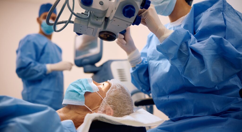
During cataract surgery, maintaining steady eye pressure is critical. Surgeons use advanced techniques and modern phacoemulsification machines to control fluid dynamics precisely. Careful pressure management minimises sudden fluctuations, protecting the optic nerve throughout the operation. This approach contributes significantly to both safety and long-term visual outcomes.
Strategies for safe cataract surgery in angle-closure glaucoma
- Adjustment of glaucoma medications pre-operatively
- Use of laser iridotomy when indicated
- Precise fluid control during surgery
- Monitoring of intraocular pressure throughout the procedure
- Protection of the optic nerve and surrounding structures
Post-operative follow-up ensures that pressure remains stable and any complications are addressed promptly. This ongoing care supports optimal recovery and long-term eye health.
Recovery and Ongoing Monitoring

Recovery after cataract surgery is usually smooth for most patients. Vision often improves quickly, and discomfort is generally minimal. Eye pressure is monitored closely in the early post-operative period to ensure stability. Follow-up visits may be more frequent to address any changes promptly.
Long-term monitoring remains essential even after successful surgery. Cataract removal improves optical clarity but does not eliminate glaucoma. Regular assessments help ensure the optic nerve remains stable over time. Ongoing care supports long-term visual health and confidence.
Key aspects of post-operative care include:
- Regular eye pressure checks in the early recovery phase
- Continued use of prescribed glaucoma medications
- Scheduled follow-up appointments and testing
- Monitoring for pressure spikes or inflammation
- Long-term optic nerve and visual field assessment
Visual and Quality-of-Life Benefits

Removing a cataract improves visual clarity, contrast, and colour perception. Many people notice that everyday activities become easier and more comfortable. Vision often feels sharper and less hazy. This improved clarity allows the eyes to work more efficiently in daily situations.
Practical tasks frequently become more manageable after surgery. Reading small print, driving in low light, and recognising faces often improve. Reduced glare and better contrast can make visual tasks less tiring. These changes contribute to a more reliable and functional visual experience.
When combined with stable eye pressure, these visual improvements often increase confidence. Better vision supports independence and social engagement. Activities that once felt difficult may feel achievable again. This positive change can also enhance emotional wellbeing and overall quality of life.
Common Benefits Patients Notice
- Clearer, sharper vision
- Improved contrast and colour perception
- Easier reading and screen use
- Greater confidence with driving and mobility
- Enhanced independence in daily activities
FAQs:
1. Can cataract surgery help manage angle-closure glaucoma?
You can benefit from cataract surgery because removing the thickened lens often widens the drainage angle. We know this helps fluid leave the eye more efficiently. Eye pressure becomes easier to control afterwards. This can protect your optic nerve and support long-term vision.
2. Why does cataract surgery improve eye pressure in angle-closure glaucoma?
You should understand that a thicker lens pushes the iris forward, narrowing fluid drainage. By replacing it with a thin artificial lens, we create more space in the front of the eye. This helps fluid flow naturally. The result is often more stable eye pressure and fewer spikes.
3. Is cataract surgery recommended even if your vision seems fine?
You may still benefit from early surgery. We sometimes suggest it to prevent future pressure problems rather than simply restore sight. This proactive approach reduces risk to the optic nerve. Early intervention can maintain long-term eye health.
4. How do acute and chronic angle-closure glaucoma differ?
You should know acute attacks happen suddenly with pain, blurred vision, and nausea. Chronic angle closure develops gradually and may have few or no symptoms. We emphasise early detection in both cases to prevent permanent damage. Regular check-ups are crucial.
5. Will cataract surgery reduce your need for glaucoma medications?
You might need fewer drops afterwards, but this varies between patients. We monitor your pressure closely after surgery. Some experience more stable readings, reducing medication dependence. Consistent follow-up ensures your eye remains protected.
6. What pre-surgical assessments are important for you?
You will undergo thorough evaluation before surgery. We examine the drainage angle, optic nerve, and overall eye structure. Visual field testing sets a baseline for comparison. This careful preparation helps plan a safe and effective procedure.
7. How is surgery performed safely for angle-closure glaucoma?
You should know that maintaining steady eye pressure during surgery is essential. We use modern equipment and refined techniques to control fluid dynamics. Sudden fluctuations are minimised. This protects your optic nerve throughout the operation.
8. What should you expect during recovery?
You can generally expect smooth recovery with improved vision. We monitor eye pressure closely in the early weeks. Discomfort is usually minimal. Follow-up visits ensure your eyes remain stable and any changes are addressed quickly.
9. Will cataract surgery restore lost vision from glaucoma?
You should understand that existing glaucoma damage cannot be reversed. We focus on improving clarity by removing the cataract. This makes remaining vision more functional. Early detection and pressure control remain essential to prevent further loss.
10. How can you maintain long-term eye health after surgery?
You play an active role by attending all follow-ups and using prescribed medications correctly. We recommend regular monitoring to detect any pressure changes. Healthy lifestyle choices support optic nerve protection. Together, these steps preserve your vision and independence.
Final Thoughts on Angle-Closure Glaucoma and Cataract Surgery
Angle-closure glaucoma is closely linked to eye anatomy, which is why cataract surgery can play such a vital role in management. By removing the thickened natural lens, the drainage angle can widen and pressure risk can be reduced.
In many cases, surgery protects vision as much as it improves clarity. With careful planning, outcomes are often very positive. If you are concerned about your glaucoma and whether cataract surgery could help manage it, get in touch with us at London Cataract Centre.
References:
- Azuara-Blanco, A., Burr, J.M., Ramsay, C.R., et al. (2016) Effectiveness of early lens extraction for the treatment of primary angle-closure glaucoma (EAGLE): a randomised controlled trial. The Lancet, 388(10052), pp.1389–1397. https://pubmed.ncbi.nlm.nih.gov/27707497/
- Brown, R.H., Camp, A.J. and Stewart, B. (2014) Reduced intraocular pressure after cataract surgery in patients with narrow angles and chronic angle-closure glaucoma. Journal of Cataract & Refractive Surgery, 40(10), pp.1610–1614. https://pubmed.ncbi.nlm.nih.gov/25134991/
- Fang, C.E.H., Mercer, R., Henein, C. and Mathew, R.G. (2021) Effectiveness in Angle-closure Glaucoma of Lens Extraction (EAGLE): clinical implications. Eye, 35, pp.2524–2526.https://www.nature.com/articles/s41433-021-01436-x
- Tanuj, D. and Friedman, D.S. (Review) (2020) Clear lens extraction in eyes with primary angle closure and primary angle-closure glaucoma. Survey of Ophthalmology, 65(6), pp.662–674.Link: https://www.sciencedirect.com/science/article/abs/pii/S0039625720300692
- Friedman, D.S. and Vedula, S.S. (2015) Lens extraction for chronic angle-closure glaucoma. Cochrane Database of Systematic Reviews, CD005555, showing evaluation of lens extraction as a treatment modality in chronic primary angle-closure glaucoma.Link: https://pmc.ncbi.nlm.nih.gov/articles/PMC4438535/