")

If you or someone you care for is living with amyloidosis and facing cataract surgery, you’re probably wondering how the condition might affect the procedure — and the healing process afterwards. That’s a fair concern. Amyloidosis isn’t just a systemic condition; when it affects the eyes, it brings its own set of unique challenges for surgeons. The changes it causes in the ocular tissues — especially the conjunctiva, sclera, and lens capsule — mean that cataract surgery can’t follow the typical path.
In this article, we’ll walk you through exactly what amyloidosis does to the eye, what it means for cataract surgery, and what both surgeons and patients should keep in mind before, during, and after the procedure.
Understanding Amyloidosis and the Eye
Amyloidosis is a rare condition in which abnormal protein deposits (amyloid) accumulate in tissues throughout the body. These deposits can disrupt normal tissue architecture and function — and the eyes are no exception. While systemic amyloidosis most often targets organs like the heart, kidneys, and liver, ocular involvement occurs more frequently than many realise, especially in advanced disease or certain types like AL (light chain) amyloidosis.
In the eye, amyloid deposits can affect several structures, including:
- Conjunctiva: leading to fragility, spontaneous haemorrhage, and abnormal healing.
- Lacrimal gland and canaliculi: resulting in dry eye and epiphora.
- Orbit and extraocular muscles: sometimes causing proptosis or restricted movement.
- Lens capsule and zonules: compromising structural support during cataract extraction.
Unlike some ocular diseases that have localised effects, amyloidosis affects tissue integrity at a fundamental level. This means that tissues don’t just become fragile — they behave differently in response to trauma, including surgical trauma. That changes the game completely for cataract surgery.
Recognising Conjunctival Fragility in Amyloidosis
One of the most distinctive ocular signs in amyloidosis is increased conjunctival fragility. In many patients, even minimal manipulation — like placing a speculum or administering anaesthetic drops — can result in sub-conjunctival haemorrhages or even microperforations. This fragility stems from amyloid infiltration into the conjunctival stroma and vessel walls, weakening the structural integrity.
For cataract surgery, this presents a few direct issues:
- Surgical access: Widening the palpebral fissure or rotating the globe during surgery can result in conjunctival tearing or bleeding.
- Topical anaesthesia: Repeated drop instillation may irritate the fragile surface, and subconjunctival injections are often avoided altogether.
- Post-operative healing: The conjunctiva may heal more slowly or abnormally, increasing the risk of infection or scarring.
To address this, many surgeons modify their pre-op and intra-op routines. For example, they may use lubricated eyelid retractors rather than metal specula, avoid aggressive conjunctival manipulation, and switch to intracameral anaesthesia.
How Amyloid Affects the Lens Capsule and Zonules
The crystalline lens sits behind the iris, held in place by fine fibres called zonules, which anchor it to the surrounding ciliary body. During cataract surgery, the front portion of the lens capsule is opened (a step called capsulorhexis), and the cloudy lens is emulsified and replaced with an artificial lens implant.
But in amyloidosis, this step becomes significantly more delicate.
Amyloid deposits can:
- Weaken zonular fibres, making them prone to rupture during lens manipulation.
- Infiltrate the capsule, making it stiffer and less elastic.
- Reduce visualisation, especially if the capsule becomes opacified or irregular due to protein deposition.
These changes raise the risk of complications such as capsular tears, dropped lens fragments, and poor support for the intraocular lens (IOL). Surgeons may need to be prepared with backup strategies — such as using capsular tension rings or anterior chamber IOLs — in case standard placement fails.
Capsule Support and IOL Selection
In eyes where zonular weakness is suspected, implanting an IOL into the capsular bag might not be safe. Instead, the surgeon might opt for alternative placements:
- Sulcus placement: positioning the lens just outside the capsular bag, though this requires intact posterior capsule support.
- Iris-claw lenses: clipped to the iris, bypassing the need for zonular support.
- Scleral-fixated IOLs: sutured to the wall of the eye, often as a last resort due to surgical complexity and risks.
Not all IOLs are created equal when it comes to patients with fragile ocular structures. Heavier, multifocal lenses may be avoided due to their reliance on precise centration and zonular stability. Instead, lightweight, monofocal lenses with proven long-term centration are often the preferred choice.
Tailoring Surgical Instruments and Techniques for Amyloid-Affected Eyes
Operating on an eye affected by amyloidosis often demands subtle but important adjustments to standard surgical tools and approaches. Even something as simple as a standard keratome incision may need to be re-evaluated if the sclera is stiffened or discoloured from amyloid deposits. Surgeons often opt for finer, sharper instruments that exert less pressure on the ocular surface, reducing the risk of micro-tears or unexpected tissue deformation. Additionally, cohesive viscoelastics may be favoured over dispersive ones to maintain chamber stability without overwhelming fragile anterior segment structures.
In some cases, amyloid infiltration makes the anterior capsule more rigid, limiting the ability to perform a smooth continuous curvilinear capsulorhexis. To counter this, some surgeons may use micro-scissors or femtosecond lasers (if available and safe in that patient) to enhance precision. During phacoemulsification, lower energy settings and gentle fluidics are often chosen to avoid hydrodissection-related rupture in already weakened capsular bags. These small changes — each based on the individual’s ocular condition — can add up to a major difference in surgical safety and outcomes.
Managing Inflammation and Post-Operative Healing

One of the underappreciated features of systemic amyloidosis is that it alters inflammatory responses. In some patients, inflammation may be blunted, while in others, it may be exaggerated due to vascular fragility and altered immune signalling.
Here’s what this means for cataract surgery recovery:
- Oedema: Amyloid infiltration can impair lymphatic and vascular drainage, leading to prolonged corneal or macular oedema.
- Haemorrhage: Even mild trauma during surgery may cause persistent bleeding, especially from the conjunctiva or scleral vessels.
- Dry eye: Involvement of the lacrimal gland or meibomian glands can worsen surface healing and increase discomfort.
Post-op care must be customised. Patients often require preservative-free lubricants, longer courses of anti-inflammatories, and close monitoring for delayed complications like cystoid macular oedema or IOL decentration.
Sedation and Anaesthetic Considerations
Systemic amyloidosis doesn’t just affect the eye — it also involves the heart, kidneys, and liver. These systems play a major role in metabolising drugs and regulating sedation safety.
Some patients may have:
- Cardiac amyloidosis (restrictive cardiomyopathy) which increases risk during conscious sedation.
- Renal involvement that alters excretion of anaesthetic metabolites.
- Autonomic neuropathy affecting blood pressure control during surgery.
Because of this, local anaesthesia with minimal sedation is generally preferred. When deeper sedation is necessary, close collaboration with an anaesthetist experienced in managing amyloidosis is critical.
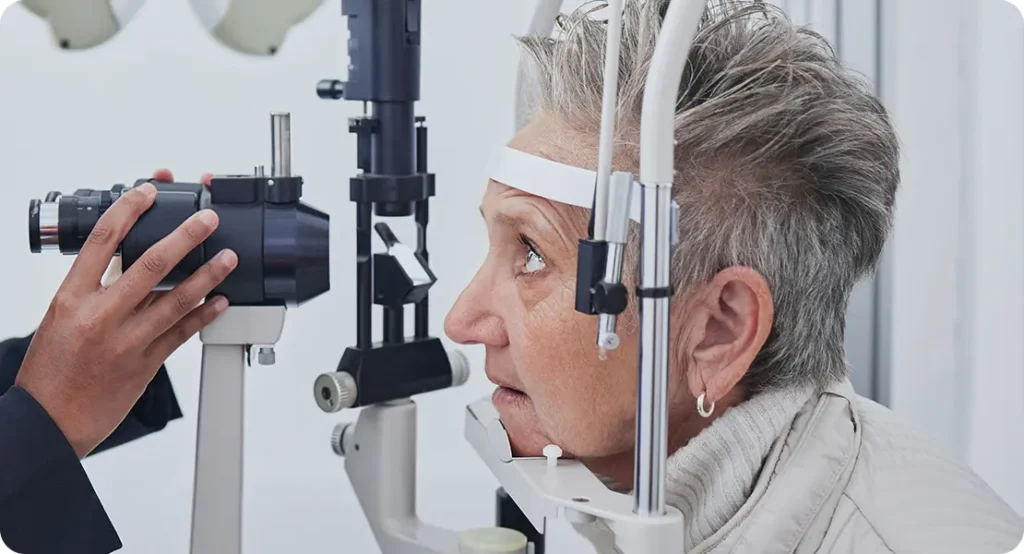
The Role of Preoperative Evaluation
Thorough preoperative assessment is crucial when planning cataract surgery in patients with amyloidosis. A standard cataract evaluation won’t be enough — here’s what needs to be added:
- Slit lamp assessment of conjunctival and scleral integrity.
- Ultrasound biomicroscopy (UBM) or anterior segment OCT to assess zonular support.
- OCT macula to rule out pre-existing macular oedema.
- Cardiology clearance, particularly in AL amyloidosis with suspected cardiac involvement.
By taking a more detailed approach upfront, surgeons can better plan the type of IOL to use, the anaesthetic technique, and potential postoperative needs.
Setting Realistic Expectations with Patients
Patients living with amyloidosis are often used to dealing with complex health concerns. But even so, it’s important to make sure expectations around cataract surgery outcomes are managed sensitively and honestly.
Here’s what to communicate clearly:
- The surgery may be technically more difficult than usual due to tissue fragility.
- Vision may not improve as dramatically if the retina or macula has pre-existing issues.
- There’s a higher chance of needing additional procedures — either for IOL repositioning, prolonged inflammation, or dry eye management.
When expectations are realistic, and both patient and surgeon are on the same page, satisfaction with the outcome tends to be higher.
Special Cases: Localised Ocular Amyloidosis
In some rare cases, amyloidosis may be isolated to the eye — such as primary conjunctival or vitreous amyloidosis. These are separate from systemic forms and have different surgical implications.
- Conjunctival-only amyloidosis may limit exposure or make incisions more difficult.
- Vitreous amyloid (involving the jelly at the back of the eye) can cause floaters and might require vitrectomy alongside or before cataract surgery.
For these patients, co-management with a retinal specialist or ocular pathologist may be needed.
Post-Operative Monitoring and Follow-Up
The post-op period in patients with amyloidosis should be handled proactively, with early detection of complications the top priority.
Key elements of follow-up include:
- Close pressure monitoring — amyloid deposits may interfere with outflow, increasing glaucoma risk.
- Early OCT imaging to catch subtle macular oedema before symptoms develop.
- Frequent surface checks to prevent infections from poorly healing conjunctival wounds.
If issues do arise, don’t panic. With early intervention, most complications can be managed effectively.
Psychological Considerations and Support for Patients with Rare Systemic Conditions
Living with a rare systemic condition like amyloidosis can be emotionally taxing — and when it comes time for surgery, patients often bring with them a heightened level of anxiety or fear. Many have already undergone complex medical interventions, and the idea of eye surgery may feel daunting, especially with the added uncertainty of how their condition might affect the outcome. It’s crucial for the care team to acknowledge these emotional layers and provide empathetic, clear, and consistent communication throughout the process.
Setting aside time to explain the surgical plan, backup strategies, and follow-up expectations can help reduce anxiety and improve cooperation. Written handouts or short video explanations tailored to patients with systemic disease can also support understanding. Where appropriate, involving family members or carers in pre-op discussions ensures a more supported recovery. Above all, reinforcing that the surgical team is experienced with complex cases and has a proactive management plan can make a significant difference in patient confidence and overall satisfaction.
FAQs
- Is it safe to have cataract surgery with systemic amyloidosis?
Yes, cataract surgery can be performed safely in patients with systemic amyloidosis, but it requires extra care due to the fragility of ocular tissues and possible involvement of other organs. Surgeons typically adapt their techniques and anaesthesia choices to reduce the risk of bleeding and delayed healing. - Will the surgery take longer than usual?
It often does, as surgeons take additional time to handle delicate conjunctiva, assess zonular stability, and proceed cautiously during lens removal and IOL placement. The focus is on safety and precision rather than speed, especially when amyloid infiltration affects support structures. - Can I get a multifocal lens implant?
Multifocal lenses are generally avoided in amyloidosis patients because they require excellent centration and stable capsule support, which may not be reliable in fragile or infiltrated eyes. A monofocal IOL is typically a safer and more predictable option in these cases. - What type of anaesthesia will be used?
Local anaesthesia with light sedation is the preferred approach, especially if the patient has cardiac or renal involvement from amyloidosis. General anaesthesia may be used in special circumstances but carries more risk due to systemic disease. - How soon will I notice improvement in vision?
Most patients begin to see improvements within a few days, although visual recovery can be slower than average if inflammation, dry eye, or postoperative oedema are present. Consistent follow-up is important to ensure any complications are promptly addressed. - Are there higher risks of complications?
Yes, there is a slightly higher risk of issues such as bleeding, capsular instability, delayed healing, and post-operative inflammation. However, these risks can be managed effectively with customised surgical planning and careful postoperative monitoring. - Can amyloidosis affect my other eye later?
If the underlying condition is systemic, it’s possible that both eyes will develop cataracts or amyloid-related changes over time. Each eye should be assessed separately, and surgery should be timed based on vision needs and overall health. - Will I need additional medications after surgery?
You may need a longer course of anti-inflammatory drops, preservative-free lubricants for surface healing, and sometimes oral medications if inflammation is prolonged. Your regimen will be adjusted based on how your eye responds post-surgery. - How often will I need follow-up visits?
Patients with amyloidosis typically need more frequent follow-up visits in the first few weeks to check for complications such as increased eye pressure, inflammation, or IOL decentration. Your ophthalmologist will decide the schedule based on your recovery. - Should I stop my amyloidosis medications before surgery?
This depends on the type of medication and your overall health. Some drugs may affect healing or bleeding risk, but you should never stop any systemic treatment without first consulting both your ophthalmologist and your treating physician.
Final Thoughts
Cataract surgery in patients with amyloidosis isn’t something to be feared — but it does require care, planning, and a different approach from the usual. If you’re dealing with this condition, make sure you’re working with a cataract surgeon who understands the nuances of systemic disease and has experience adapting techniques to match the eye’s condition.
At the London Cataract Centre, we take pride in handling complex cataract cases, including those affected by rare conditions like amyloidosis. We’re here to guide you through every step of the process — from pre-operative planning to your final follow-up.
References
- Suesskind, D., Ziemssen, F. & Rohrbach, J. M., 2015. Conjunctival amyloidosis – clinical and histopathologic features. Graefe’s Archive for Clinical and Experimental Ophthalmology, 253(8), pp.1377–1383. Available at: https://pubmed.ncbi.nlm.nih.gov/25619666/
- Ocular Amyloidosis, 2024. StatPearls. Available at: https://www.ncbi.nlm.nih.gov/books/NBK603727/
- Falk, R. H., Alexander, K. M., Liao, R. & Dorbala, S., 2016. AL (Light‑Chain) Cardiac Amyloidosis: A Review of Diagnosis and Therapy. Journal of the American College of Cardiology, 68(12), pp.1323–1341. Available at: https://pubmed.ncbi.nlm.nih.gov/27634125/
- Casado, G. et al., 2015. Systemic amyloidosis with bilateral conjunctival involvement: a case report. BMC Ophthalmology, 15, 91. Available at: https://bmcophthalmol.biomedcentral.com/articles/10.1186/s12886-015-0075-2
- Seman, S. & Salti, H., 2018. Amyloidosis and ocular involvement: an overview. Ophthalmology and Therapy, 7(2), pp. 243–261. (Although the PubMed entry for this article uses PMID 31829761, the full text is accessible via PubMed.) Available at: https://pubmed.ncbi.nlm.nih.gov/31829761/