")

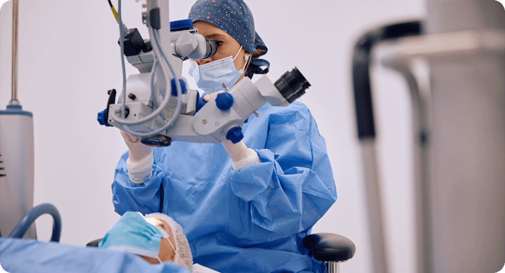
When you think of cataract surgery, you might imagine a fairly straightforward procedure—pop out the cloudy lens, pop in a clear one, and job done. And for many people, that’s pretty much the case. But not all cataract surgeries are created equal. For patients with coexisting eye conditions—like corneal irregularities, glaucoma, macular degeneration, or previous surgeries—the approach needs to be a lot more personalised.
That’s where preoperative imaging really earns its stripes. Technologies like optical coherence tomography (OCT) and Scheimpflug imaging (Pentacam being the most well-known) allow surgeons to see what the naked eye can’t. They uncover risks, clarify the anatomy, and ultimately guide safer, smarter surgical decisions.
In this article, we’ll explore why preoperative imaging matters so much in complex cataract cases, how OCT and Pentacam work, and how they shape everything from intraocular lens (IOL) selection to surgical technique.
Why Complex Cataract Cases Demand More Than a Basic Workup
Let’s start with the obvious: cataract surgery is a highly successful and low-risk procedure—when everything else in the eye is normal. But as soon as you throw another condition into the mix, the predictability of the outcome can wobble.
Patients with irregular corneas, shallow anterior chambers, dense posterior polar cataracts, epiretinal membranes, or previous laser surgeries have more variables in play. These aren’t eyes you want to approach with assumptions. Imaging steps in as a diagnostic safety net.
Traditional slit-lamp exams and ultrasound can only get you so far. OCT and Pentacam fill in the blanks by revealing the integrity of the macula, the shape of the cornea, anterior chamber depth, and more. Without that information, you’re essentially flying blind—and that’s a risk no surgeon wants to take.
OCT: Seeing Below the Surface of the Retina
OCT is a game-changer in complex cataract planning. Think of it as ultrasound using light instead of sound. It creates high-resolution, cross-sectional images of the retina and anterior segment, offering a wealth of information before you ever pick up a scalpel.
This is especially important in patients with subtle or undiagnosed macular conditions. Sometimes the cataract is so dense it hides what’s going on behind it. But in other cases, OCT uncovers pathology like macular oedema, epiretinal membranes, or dry age-related macular degeneration that can influence IOL choice or even postpone surgery.
Why does this matter? Because a patient with a compromised macula won’t benefit from a premium multifocal IOL—and placing one could lead to disappointment, even litigation. OCT helps manage expectations and match technology to anatomy.
Anterior Segment OCT: The Unsung Hero in Surgical Planning
While retinal OCT gets a lot of the limelight, anterior segment OCT (AS-OCT) deserves equal attention in tricky cases. It provides detailed imaging of the cornea, angle, iris, and anterior lens capsule.
In patients with narrow angles, for example, AS-OCT can help confirm the degree of angle closure and whether laser peripheral iridotomy should be done before surgery. In trauma cases or those with weak zonules, AS-OCT can detect subtle phacodonesis and capsule instability, guiding whether capsular tension rings or other stabilisers might be needed.
It’s also valuable in eyes with previous refractive surgery. The corneal curvature may not tell the full story, but anterior segment OCT can reveal interface issues or residual flap edges that might influence your incision planning or affect corneal healing.
Pentacam: Mapping the Eye in Three Dimensions
Now, let’s talk about Pentacam. This device uses Scheimpflug imaging—a technique that captures sharp images of the anterior segment by rotating a camera around the eye. It’s especially powerful for evaluating the cornea and lens in 3D.
In keratoconus or post-LASIK eyes, Pentacam reveals topographic abnormalities that can throw off IOL calculations. You can’t trust standard keratometry in those cases. Pentacam gives you curvature maps, pachymetry data, posterior corneal astigmatism, and elevation details that are essential for accurate lens power estimation.
The software also helps you simulate outcomes—so you can predict how a toric lens might behave or whether an aspheric IOL would give better contrast in a particular patient. For irregular corneas, it can flag whether a scleral fixated IOL or a staged corneal procedure might be the safer route.
Identifying Subclinical Corneal Pathology
One of the greatest dangers in cataract surgery is operating on an eye you think is normal. Subclinical keratoconus, forme fruste dystrophies, and early Fuchs’ dystrophy can look deceptively stable on slit-lamp exam.
Pentacam helps you catch these landmines before they explode. If the corneal map shows inferior steepening, central thinning, or high posterior elevation, you’ll want to reconsider implanting a multifocal lens. These eyes may decompensate post-op and end up worse than before surgery.
Likewise, early endothelial cell loss patterns on Pentacam pachymetry can push you to opt for a less aggressive fluidic setting or even schedule a staged DMEK later. The imaging doesn’t just inform; it redirects the entire surgical roadmap.
The Role in Posterior Polar Cataracts
If you’ve ever handled a posterior polar cataract, you’ll know the risks. These are fragile lenses with a notorious tendency for capsular rupture. Traditional imaging doesn’t always detect the extent of posterior capsule weakness.
That’s where anterior segment OCT steps up. It can sometimes identify thinning or pre-existing breaches in the posterior capsule. And even if it can’t, the knowledge of this being a posterior polar case, confirmed by characteristic imaging features, encourages a no-hydrodissection approach and altered phaco technique.
Planning with imaging, in this case, may prevent one of the most dreaded complications in cataract surgery—a dropped nucleus. In high-stakes eyes, information really is power.
Post-Refractive Surgery Eyes: A Calculative Nightmare
Cataract patients who’ve had LASIK, PRK, or RK are increasingly common. These eyes defy the assumptions baked into standard IOL formulae. The altered corneal shape throws off your anterior and posterior curvature readings, making the IOL prediction a guessing game—unless you bring imaging into the equation.
Pentacam offers true net power readings and posterior corneal curvature analysis, allowing for better use of formulas like Barrett True-K or Haigis-L. Combining Pentacam data with modern calculators often yields significantly better outcomes than traditional methods.
Without this imaging, you’re often undercorrecting or overcorrecting—leaving the patient with frustration and more procedures. Preoperative imaging brings the guesswork down to a minimum and the predictability up to modern expectations.
When Glaucoma Is in the Picture
Patients with glaucoma bring unique risks. Intraoperative pressure spikes, medication toxicity, and angle status all influence your planning. OCT and Pentacam together provide a layered understanding of both anterior and posterior anatomy.
With OCT, you can check for nerve fibre layer loss and ganglion cell thinning. These findings might make you reconsider the use of premium lenses, as contrast sensitivity is already impaired. Pentacam, meanwhile, helps confirm angle status and anterior chamber depth—especially important when using phaco techniques that increase fluid turbulence.
In narrow angles or chronic angle closure, this imaging could be the reason you choose to perform a combined cataract and MIGS (minimally invasive glaucoma surgery) procedure. Without it, you may miss the opportunity for a more comprehensive surgical plan.
The Impact on IOL Selection
Premium IOLs—like toric, multifocal, and extended depth of focus lenses—are not a one-size-fits-all solution. Their success depends entirely on the integrity of the cornea, the stability of the macula, and the predictability of surgical outcomes.
Preoperative imaging helps you avoid missteps. If the cornea is irregular, the retina is compromised, or the ocular surface is unstable, you can guide the patient toward a monofocal IOL with realistic expectations. That decision, made early and with clarity, can prevent post-op disappointment.
And in appropriate cases, the imaging helps you justify using premium lenses. It provides the evidence that conditions are optimal—and supports your recommendation with data, not just intuition.
Imaging for Surgical Strategy and Technique
Beyond choosing the lens, imaging also influences your surgical approach. Shallow anterior chambers or dense cataracts might push you toward femtosecond laser-assisted techniques. Eyes with weak zonules might warrant capsule tension rings or modified nucleus chopping.
Pentacam and OCT data can also flag when a pupil expansion device might be needed or whether intraoperative OCT might be helpful. These aren’t just diagnostic tools—they’re strategy setters. They shape how you enter the eye, how you navigate within it, and how you close.
Streamlining Post-Op Monitoring
Even after the cataract is out, these imaging tools continue to be useful. OCT allows you to monitor the macula for cystoid macular oedema or fluid accumulation, especially in diabetic or uveitic eyes. Pentacam, meanwhile, helps track corneal healing and detect subtle signs of decompensation in high-risk cases.
Imaging isn’t just a pre-op luxury—it’s a longitudinal investment in quality care.
FAQ: Preoperative Imaging in Complex Cataract Cases
1. Why isn’t a basic eye exam enough before cataract surgery?
A standard eye exam can pick up many things—like visual acuity, lens opacity, and pressure—but it doesn’t give a full picture of what’s happening beneath the surface. When dealing with complex cases, such as eyes with previous laser surgery, subtle macular disease, or corneal irregularities, you need to go deeper. That’s where preoperative imaging comes in. Technologies like OCT and Pentacam allow for precise visualisation of the retina, cornea, anterior chamber, and lens capsule—giving your surgeon the kind of data a torchlight exam simply can’t.
Skipping advanced imaging in a complex case is a bit like planning a road trip without checking the weather or the route—it increases the risk of surprises. For example, undiagnosed macular pathology could ruin the visual outcome if a premium IOL is placed. Similarly, corneal mapping might uncover irregularities that contraindicate toric lenses or suggest a need for surgical adjustment.
So while basic tests are still useful, they are often insufficient in ensuring the best possible results. If your case isn’t textbook straightforward, imaging gives your surgeon the confidence to choose the right lens and the safest surgical plan for your eyes.
2. What exactly does OCT show that other tests don’t?
OCT (Optical Coherence Tomography) provides cross-sectional, high-resolution images of the eye’s internal structures. Most importantly, it reveals the health of the macula and optic nerve—two areas crucial for good vision. While conditions like epiretinal membranes, macular degeneration, and diabetic macular oedema can sometimes be suspected from symptoms or basic tests, OCT confirms their presence with much greater clarity.
In the anterior segment, OCT also evaluates angle structure, anterior chamber depth, corneal layers, and lens position. This is particularly useful in patients with narrow angles, glaucoma, or suspected zonular weakness. Anterior segment OCT can detect subtle capsule instability or corneal pathology that may affect surgical decisions.
In short, OCT gives your surgeon both a retinal and corneal “status report.” And in borderline or complicated cases, that can mean the difference between a smooth recovery and one fraught with issues.
3. What is Pentacam, and why is it useful before cataract surgery?
Pentacam is a type of Scheimpflug imaging system that takes a 3D scan of the anterior segment of your eye. Unlike standard topography, it maps both the front and back surfaces of the cornea, providing more accurate data about curvature, thickness, and elevation. It also measures anterior chamber depth, lens density, and pupil size under different lighting conditions.
This matters because certain corneal shapes—like those seen in keratoconus or post-LASIK eyes—can dramatically affect IOL power calculations. In these patients, regular biometry is often inaccurate, and Pentacam provides the alternative metrics needed to make good surgical choices.
Additionally, Pentacam helps uncover hidden issues like corneal ectasia or early Fuchs’ dystrophy. Knowing these exist before surgery gives your surgeon the chance to modify their technique or IOL selection—and may even delay surgery until further stabilisation is achieved.
4. Is preoperative imaging only useful for people with known eye conditions?
Not at all. While it’s absolutely essential in known complex cases—like those with macular disease, prior refractive surgery, or glaucoma—it’s also valuable in cases where problems haven’t yet been diagnosed. Some people have subclinical issues that wouldn’t be caught without imaging.
For example, early keratoconus or mild macular oedema might be completely missed on routine exam but picked up on imaging. If left undetected, these could lead to poor visual outcomes or the wrong type of IOL being implanted. In some cases, imaging changes the course of treatment entirely—prompting a different type of lens, a combination surgery, or even a decision to delay the procedure.
So even if you’re not aware of having any additional eye issues, preoperative imaging acts like a pre-flight safety check—confirming that everything is in good condition before take-off.
5. Should I ask my surgeon if they use OCT and Pentacam before surgery?
Yes—especially if your eyes are anything other than completely straightforward. If you’ve had laser eye surgery in the past, have a history of diabetes, glaucoma, or notice changes in your central vision, asking about preoperative imaging is not just appropriate—it’s smart.
A good cataract surgeon should already be incorporating these technologies into their standard workflow for complex cases. But confirming that they do so also gives you peace of mind. It shows they’re going beyond basic diagnostics and investing in technologies that reduce surgical risk and improve visual outcomes.
You’re trusting someone with your eyesight. It’s perfectly reasonable to want to know they’re using the best tools available.
Final Thoughts
Cataract surgery is no longer a one-size-fits-all procedure—especially not in eyes with coexisting pathology. The days of relying solely on slit-lamp exams and A-scan ultrasound are over. With tools like OCT and Pentacam, surgeons can now tailor their approach with precision, reduce risks, and vastly improve patient outcomes.
If you’re considering private cataract surgery in London, make sure your surgeon incorporates these advanced imaging modalities into their planning. It’s not just about removing a cloudy lens—it’s about restoring your vision with accuracy, confidence, and the highest standard of care.
References
- Zhang, T., Zhu, X. and He, W., 2020. Application of anterior segment optical coherence tomography in cataract surgery. Eye and Vision, 7(1), pp.1–11. https://doi.org/10.1186/s40662-020-00182-7
- Belin, M.W., Khachikian, S.S. and Ambrosio, R., 2011. Validation of the Pentacam Belin/Ambrósio Enhanced Ectasia Display: A New Method for Detecting Ectatic Corneal Disease. Journal of Refractive Surgery, 27(10), pp.803–810. Available at: https://www.healio.com/ophthalmology/journals/jrs
- Day, A.C., Gore, D.M., Bunce, C. and Evans, J.R., 2016. Laser-assisted cataract surgery versus standard ultrasound phacoemulsification cataract surgery. Cochrane Database of Systematic Reviews, Issue 7. Available at: https://www.cochranelibrary.com
- Hirnschall, N., Amir-Asgari, S., Maedel, S. and Findl, O., 2013. Predicting Postoperative Refraction in Eyes With Previous Myopic LASIK Using Scheimpflug Imaging and Ray-Tracing Calculations. Journal of Cataract and Refractive Surgery, 39(9), pp.1347–1352.
- Tsorbatzoglou, A., Sidiropoulos, G., Moschos, M.M. and Gatzioufas, Z., 2018. Optical coherence tomography findings in posterior polar cataract: A case series. American Journal of Ophthalmology Case Reports, 10, pp.35–38. Available at: https://doi.org/10.1016/j.ajoc.2018.01.009