")

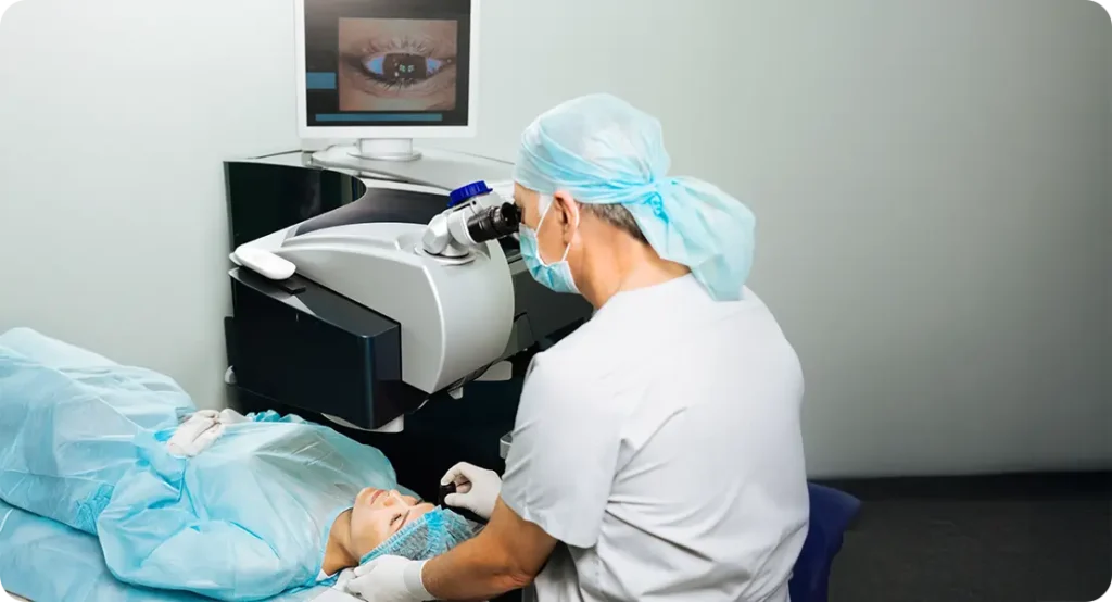
If you’re getting ready for cataract surgery or simply curious about how we’re achieving better and better post-op results, you’ll want to know what’s happening behind the scenes. And a big part of that involves optical biometry. This technology has evolved dramatically over the past decade, playing a crucial role in how we determine the power of the intraocular lens (IOL) implanted during cataract surgery.
But the story doesn’t stop there. New research is taking things even further—using artificial intelligence and more detailed biometric data to refine these predictions. The aim? To make post-surgery vision sharper, more predictable, and tailored to your eyes like never before.
What Is Optical Biometry, Really?
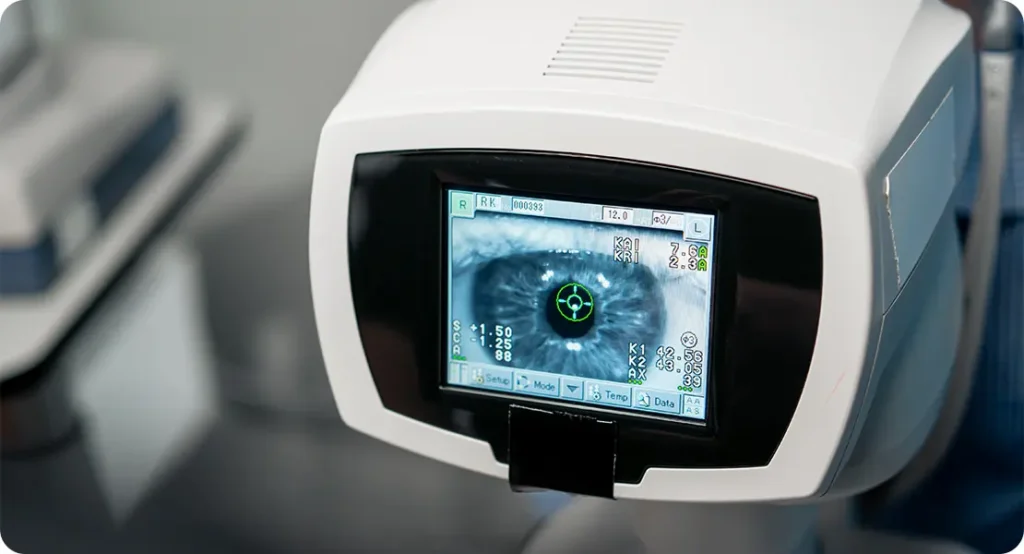
Optical biometry is the gold standard for measuring the eye’s dimensions before cataract surgery. It’s a quick, contactless procedure that captures incredibly precise data points about your eye. Think of it as a digital map of your unique eye structure, helping your surgeon calculate exactly which lens power will give you the best possible vision after surgery. Instead of just estimating or relying on traditional tools, optical biometry brings scientific accuracy to the process, turning IOL selection into a personalised decision rather than a one-size-fits-all choice.
The main measurements taken during optical biometry include axial length (the distance from the front to the back of your eye), anterior chamber depth (the space between the cornea and the lens), corneal curvature (how steep or flat your cornea is), and lens thickness. Some machines also measure the “white-to-white” distance, which is the visible horizontal diameter of the iris. These figures are plugged into lens power formulae that predict what strength of IOL will bring your vision closest to normal. It’s these calculations that can make the difference between needing glasses post-op—or not.
What makes optical biometry superior to older techniques like ultrasound is the level of detail and repeatability. Ultrasound biometry required physical contact with the eye and relied heavily on the skill of the technician. Optical methods, particularly those using partial coherence interferometry (PCI) and swept-source optical coherence tomography (SS-OCT), are faster, more comfortable for the patient, and more reliable from one test to the next. That means less chance of human error and more confidence in the surgical plan.
Accuracy in optical biometry isn’t just a technical point—it has real-world consequences. If the axial length is off by even a small fraction of a millimetre, the predicted lens power can be significantly wrong. This could leave patients with unexpected hyperopia (farsightedness) or myopia (nearsightedness) after surgery. That’s why modern cataract surgery planning hinges on high-quality biometric data. With the right measurements, patients have a much better chance of achieving 20/20 or near-perfect results without needing corrective lenses afterwards.
The Move from Classic to Cutting-Edge Devices
When optical biometry first emerged, it was already a major leap forward compared to ultrasound. Devices like the IOLMaster 500 set new standards by using light instead of sound to measure axial length and other vital distances. The big selling point was its non-contact approach—safer, cleaner, and more comfortable. However, it still had its weaknesses, particularly when dense cataracts made it hard for light to penetrate the eye clearly. That’s where newer technology stepped in to push boundaries even further.
The latest wave of devices, such as the IOLMaster 700 and Lenstar LS 900, utilise swept-source optical coherence tomography (SS-OCT). Unlike earlier models, these can penetrate through denser opacities and image more layers of the eye. The SS-OCT technology allows for a complete B-scan image of the eye’s cross-section, which not only helps in measurements but also lets surgeons assess the overall eye structure in much greater detail. With this level of visualisation, identifying anomalies or confirming the health of the ocular structures becomes a seamless part of the pre-operative workflow.
What sets these advanced devices apart is their software. Instead of simply providing raw measurements, they come loaded with a range of IOL power calculation formulae, including traditional models and newer AI-powered ones. Surgeons can choose from an expanded toolkit and tailor their calculations based on the patient’s specific eye anatomy and needs. Additionally, many of these devices include toric IOL calculators for astigmatism correction, and some even provide guidance on lens orientation and alignment, especially useful for premium lens options.
Perhaps most importantly, the newer devices now include the ability to measure the posterior corneal curvature—something earlier machines didn’t account for. This back surface of the cornea can subtly influence total corneal power and affect the accuracy of IOL predictions, especially for patients with astigmatism. By factoring in posterior data, the margin of error is significantly reduced. This development shows how biometry isn’t just evolving—it’s getting smarter and more complete with every generation.
Beyond Standard Measurements: Adding More Data
Traditionally, most IOL power calculations relied on just a few core variables: axial length, anterior chamber depth, and corneal curvature. These basics formed the foundation of every pre-op assessment. But as outcomes became more refined and patient expectations grew, researchers and clinicians began asking: what if we included more data? It turns out that variables like lens thickness, corneal diameter (white-to-white), and even pupil size can subtly influence lens positioning and, ultimately, visual outcomes.
Modern biometers have responded by making these additional measurements routine. Devices like the Pentacam AXL and OA-2000 now offer full anterior segment scans, giving a 3D view of the front of the eye. Others, like the Argos SS-OCT, go further by combining posterior segment imaging as well. This means surgeons now have access to a complete anatomical profile, not just numbers on a chart. It’s the difference between seeing a few key data points and understanding the full geometry of the eye in context.
But gathering more data is only half the story. The real breakthrough has come from how that data is interpreted. With so many variables at play, even experienced surgeons can struggle to know which ones matter most in a given case. That’s where artificial intelligence and machine learning come in. These systems can process massive datasets—thousands of eyes, surgeries, and outcomes—to uncover patterns no human could reasonably detect. This allows for far more personalised and accurate lens predictions.

Adding these extra data layers has been particularly valuable in borderline or unusual cases—such as very long eyes, post-refractive surgery patients, or those with high corneal astigmatism. In these instances, the old standard measurements weren’t enough to get an accurate prediction. But with expanded biometry and machine-driven analysis, even complex eyes are being planned with greater precision. It’s not just about gathering data anymore—it’s about making sense of it in a clinically meaningful way.
The Rise of Artificial Intelligence in IOL Calculations
The application of artificial intelligence in IOL power prediction has revolutionised how we approach cataract surgery planning. Instead of relying solely on rigid mathematical models like SRK/T or Hoffer Q, we now have access to AI-based algorithms that adapt and learn over time. These systems analyse real-world surgical outcomes and refine their predictions continuously, improving accuracy as more data is fed into them. It’s a living, evolving tool that gets better with every case.
One of the most well-known AI models is the Hill-RBF (Radial Basis Function) calculator. It doesn’t use a traditional formula; instead, it relies on a massive dataset of real surgeries and their outcomes to identify patterns. When you input your eye measurements, the model compares them with thousands of similar eyes and tells you what lens power worked best in those cases. It’s like tapping into the experience of a global network of surgeons, condensed into one instant recommendation.
The Kane formula is another standout—it blends theoretical optics with AI-based optimisation. It combines the mathematical understanding of how lenses interact with light with machine learning insights from actual patients. This hybrid model is especially useful for atypical eyes—those with unusually short or long axial lengths, steep or flat corneas, or histories of laser surgery. Where traditional formulae might struggle, the Kane formula holds its ground with consistent, accurate results.
Perhaps the most exciting part of this AI revolution is the role of big data. Thanks to cloud-based platforms, anonymised biometric and surgical outcome data from clinics all over the world is now being pooled. These enormous datasets fuel the continual training and upgrading of prediction models. What used to be a static formula is now a dynamic system—constantly improving, learning from new cases, and helping deliver better outcomes for the next patient in line. This feedback loop between AI, surgeons, and patients is setting a new standard for cataract surgery planning.
Tackling Outliers: Improving Accuracy for Complex Eyes
One of the most frustrating challenges in cataract surgery is achieving great results in patients with unusual eye anatomies—like those who are extremely nearsighted or farsighted, or who have had previous laser vision correction.
Traditional formulae often underperform in these eyes. But newer optical biometers and AI-driven calculators are proving more effective here. For example, the Barrett Universal II and Olsen formulae account for factors like lens position prediction and corneal shape, which are critical in extreme cases.
AI models, too, have shown impressive results with complex eyes. By learning from actual post-op outcomes in similar cases, they offer personalised predictions that significantly reduce refractive surprises. In fact, some clinics now routinely use multiple formulae and average the results—or rely entirely on AI-based systems—to maximise precision.
So, even if your eye anatomy is outside the norm, chances are much higher today that your post-op vision will hit the mark.
Toric and Multifocal IOLs: A Greater Need for Precision
If you’re choosing a premium IOL—like a toric lens to correct astigmatism or a multifocal lens for distance and near vision—the margin for error shrinks. Even a minor inaccuracy in lens power or axis alignment can impact your visual quality.
That’s where optical biometry truly shines. Devices like the Cassini and IOLMaster 700 offer precise corneal mapping, including posterior astigmatism data, helping surgeons fine-tune lens selection and placement.
New AI-guided planning platforms also suggest optimal IOL alignment and rotational stability based on the patient’s individual biomechanics. It’s a level of detail that wasn’t even possible a decade ago—and it’s making a real difference in patient satisfaction, especially among those with high visual expectations.
Optimising Outcomes: Biometry Meets Surgical Technique
Even the best IOL calculation is only as good as the surgery itself. But here’s the good news: the integration of biometry data into surgical guidance systems is closing that gap.
Many modern operating microscopes and phacoemulsification systems can import biometric measurements directly, offering real-time alignment guidance during surgery. Technologies like Callisto Eye and Verion help surgeons align toric lenses with sub-degree accuracy.
Some systems even account for surgically induced astigmatism (SIA), adjusting the IOL choice or placement dynamically. It’s a seamless blend of pre-op planning and intra-op execution—all powered by data that began with optical biometry.
Biometry in Post-Operative Assessment
The role of biometry doesn’t stop after surgery. Post-op optical biometry can confirm IOL positioning, assess capsular bag stability, and detect rotation in toric lenses.
In some clinics, repeat biometry is part of routine follow-up, especially if the patient’s vision isn’t as expected. Identifying a rotated toric IOL early can mean a quick fix rather than prolonged dissatisfaction.
Moreover, post-op biometric data is feeding back into AI databases, constantly improving future predictions. This loop—from pre-op measurement to post-op verification and AI model update—is a powerful engine for ongoing improvement.
Research Trends and What the Future Holds
Looking ahead, the future of optical biometry lies in greater automation, smarter AI, and even more personalised prediction models.
Some research is exploring the use of intraoperative aberrometry—measuring the eye’s optical performance during surgery—to further refine IOL selection. Others are working on integrating biometric data with genetic markers to understand why some patients heal faster or experience different visual outcomes.
And we may even see “dynamic” IOLs on the horizon—adjustable lenses that can be fine-tuned post-operatively using light or laser. Accurate biometry will be central to programming these lenses, both before and after implantation.
There’s also growing interest in tele-biometry, where measurements are taken at local clinics or via mobile platforms and analysed centrally using cloud-based AI systems. This could bring world-class surgical planning to even the most remote areas.
Final Thoughts: The Promise of Precision
If you’re preparing for cataract surgery today, you’re benefitting from a technological revolution that’s quietly reshaping outcomes. Optical biometry has moved from being a measurement tool to the cornerstone of precision eye surgery.
With the help of AI, high-resolution imaging, and global data-sharing, IOL power prediction is no longer a best guess—it’s a personalised science. That means fewer surprises, better outcomes, and happier patients.
So the next time you hear your eye surgeon talking about “biometry,” you’ll know it’s not just tech talk. It’s the reason your post-surgery vision could be better than you ever imagined.
References
- Holladay, J.T., 2017. Intraocular lens power calculations: past, present, and future. Current Opinion in Ophthalmology, 28(4), pp.313–318.
Available at: https://journals.lww.com/co-ophthalmology/Fulltext/2017/07000/Intraocular_lens_power_calculations__past,.10.aspx - Kane, J.X., Van Heerden, A., Atik, A. and Petsoglou, C., 2017. Accuracy of 3 new methods for intraocular lens power selection. Journal of Cataract & Refractive Surgery, 43(3), pp.333–339.
Available at: https://doi.org/10.1016/j.jcrs.2016.11.032 - Hill, W., 2021. The Hill-RBF Method: Artificial Intelligence in IOL Power Calculation. Asia-Pacific Journal of Ophthalmology, 10(5), pp.451–455.
Available at: https://journals.lww.com/apjoo/Fulltext/2021/10000/The_Hill_RBF_Method__Artificial_Intelligence_in.10.aspx - Srivannaboon, S., Chirapapaisan, C., Chirapapaisan, N. and Reinstein, D.Z., 2019. Comparison of axial length, keratometry and IOL power measurements between the IOLMaster 500 and 700. Clinical Ophthalmology, 13, pp.1361–1367.
Available at: https://www.dovepress.com/comparison-of-axial-length-keratometry-and-iol-power-measurements-bet-peer-reviewed-fulltext-article-OPTH - Cooke, D.L. and Cooke, T.L., 2016. Comparison of 9 intraocular lens power calculation formulas. Journal of Cataract & Refractive Surgery, 42(8), pp.1157–1164.
Available at: https://doi.org/10.1016/j.jcrs.2016.06.017