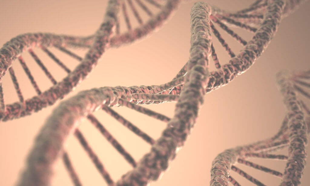
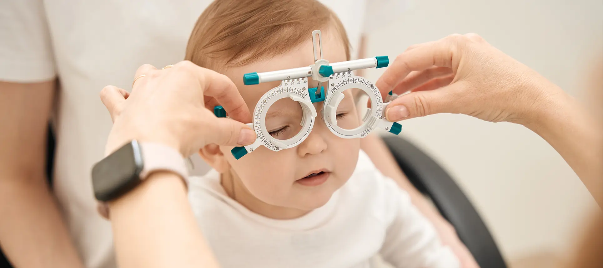
Most people think of cataracts as a condition that only affects the elderly. But what if you were born with them? Congenital cataracts—lens opacities present at birth or developing within the first year—are a different story altogether. They’re not the result of decades of UV exposure or age-related wear and tear. Instead, they often have a deeper, more complex cause: your genes.
At the heart of many of these early-onset cataracts lies a family of structural proteins known as crystallins. These are the unsung heroes that maintain the transparency and refractive power of the eye’s lens. When they’re working properly, light passes through the lens smoothly and focuses neatly on the retina. But when mutations disrupt their function—particularly in the α-, β-, or γ-crystallin groups—the lens can become cloudy, even before a baby opens their eyes for the first time.
In this article, we’re diving deep into the molecular world of crystallin proteins. We’ll explore how tiny changes in DNA can cause these crucial proteins to misfold or clump together, leading to lens opacity. And we’ll look at the latest research shaping how we diagnose, understand, and potentially treat congenital cataracts rooted in protein dysfunction.
What Are Crystallins and Why Do They Matter?
Crystallins are highly soluble, stable proteins that make up over 90% of the human lens’s dry weight. They’re not just there to take up space—they’re vital for keeping the lens clear and correctly shaped throughout life. Unlike other cells in the body, lens fibres don’t renew themselves, so the proteins within them have to last a lifetime. That’s a tall order, and it’s why stability and resistance to aggregation are so critical.
There are three main families of crystallins in the vertebrate lens:
- α-Crystallins: These are actually heat shock proteins and act like molecular chaperones. Their job is to prevent other proteins from aggregating, especially under stress conditions like oxidation or heat.
- β-Crystallins: Structurally diverse and present in multiple isoforms, β-crystallins are involved in maintaining the lens’s structural integrity.
- γ-Crystallins: These are highly stable, monomeric proteins that play a big role in the inner nucleus of the lens, especially in focusing light precisely.
Each group contributes to the overall optical properties of the lens, and their precise arrangement is key. Any disturbance in their structure or expression can disrupt the whole system—leading to lens clouding.
The Genetic Link: Inherited Mutations Behind Congenital Cataracts
A large proportion of congenital cataracts have a genetic basis, with more than 50 known genes implicated. Among these, mutations in crystallin genes (especially CRYAA, CRYAB, CRYBA1, CRYBB2, CRYGC, and CRYGD) are frequently observed. These mutations often follow an autosomal dominant inheritance pattern, meaning a child only needs one faulty copy of the gene to develop the condition.

The effects of these mutations vary. Some lead to the production of misfolded proteins that are unstable and degrade easily. Others cause proteins to aggregate into insoluble clumps that scatter light and cloud the lens. Still others interfere with the protein’s ability to interact with other lens proteins, undermining the ordered architecture of the lens fibres.
Some families have been studied for generations, revealing clear inheritance patterns and helping scientists trace the origin of specific crystallin gene mutations. Genetic testing is now becoming more common in paediatric ophthalmology, helping identify which mutation is responsible and guiding future treatment strategies.
α-Crystallins: The Molecular Chaperones
Let’s start with the α-crystallins, made up of αA- and αB-crystallin subunits. Think of them as quality control managers inside the lens. Their main job is to bind to partially unfolded or misfolded proteins and stop them from clumping together. In doing so, they maintain transparency and protein stability within the lens.
Mutations in CRYAA and CRYAB can be particularly damaging. A faulty αA-crystallin may fail to bind misfolded proteins properly, allowing them to aggregate. Alternatively, the protein itself might become unstable, creating an ironic twist—your chaperone needs a chaperone.
One well-known mutation, R116C in CRYAA, changes a single amino acid but leads to severe bilateral cataracts in infants. This substitution disrupts the protein’s structural domain, making it prone to aggregation. Another mutation, R120G in CRYAB, is associated not only with cataracts but also with myopathy, highlighting how this lens protein plays a role in other tissues too.
Recent research also suggests that these mutated α-crystallins may trigger stress responses or apoptosis (cell death) in lens epithelial cells, worsening the damage and accelerating opacification.
β-Crystallins: Structural Stability and Mutation Sensitivity
The β-crystallins are a bit more structurally complex. They form dimers or higher-order oligomers and are crucial for building the scaffolding of the lens. Mutations in β-crystallin genes like CRYBB1, CRYBB2, and CRYBA1 can destabilise these structures, leading to premature unfolding or misassembly.
One example is the W151C mutation in CRYBB2, which has been identified in families with cerulean cataracts—a distinctive form with bluish opacities. This mutation alters a key tryptophan residue essential for the correct folding of the protein, promoting aggregation and insolubility.
Another case involves CRYBA1, where a splice site mutation leads to a truncated protein missing a domain necessary for solubility. The result? A loss of function and gain of aggregation—two hits in one.
Interestingly, β-crystallins don’t work in isolation. They often form heteromers with other β-crystallins, and so a mutation in one type can disrupt the entire complex, magnifying the impact of the genetic change. This interdependency makes the system especially sensitive to mutations.
γ-Crystallins: Precision Under Pressure
The γ-crystallins are the most structurally rigid and compact of the three families. They’re monomeric, highly symmetrical, and packed tightly in the lens nucleus. Their job is to maintain precise optical density gradients without clumping or unfolding—especially important because the centre of the lens doesn’t regenerate.
Mutations in genes like CRYGC and CRYGD are strongly associated with autosomal dominant congenital cataracts. One infamous example is the P23T mutation in CRYGD, which changes a proline to a threonine. That might sound minor, but it affects protein solubility so dramatically that it precipitates out of solution, forming light-scattering aggregates.
Other mutations can affect how γ-crystallins fold. Because these proteins rely on precise β-sheet arrangements, even slight misalignments can expose hydrophobic surfaces, encouraging clumping. The T5P and R36S mutations are both examples where structural misfolding leads to insolubility.
The γ-crystallins are also vulnerable to post-translational changes like oxidation, which exacerbate the aggregation caused by mutations. This combination of genetic and environmental insults explains why some children with these mutations develop cataracts earlier or more severely than others.
Protein Misfolding and Aggregation: The Biochemical Fallout
So what’s actually happening inside the lens at the molecular level? When a crystallin protein misfolds due to a mutation, it often exposes normally hidden hydrophobic regions. These sticky surfaces attract other misfolded proteins, forming insoluble aggregates.
The problem is twofold. First, these aggregates scatter light—literally making the lens opaque. Second, they can trigger cellular stress responses. Misfolded protein accumulation in lens fibre cells can activate the unfolded protein response (UPR) or even lead to apoptosis. That’s especially damaging because lens cells, once matured, don’t regenerate.
To make matters worse, some mutations don’t just affect folding but also interfere with the protein’s interactions. For example, α-crystallin’s chaperone function can be compromised, allowing other misfolded proteins to accumulate unchecked.
Researchers have used biophysical techniques like X-ray crystallography, NMR spectroscopy, and dynamic light scattering to track how these mutations change protein structure and behaviour. In almost every case, the result is the same: a shift from soluble, transparent proteins to insoluble, scattering clumps.
How These Mutations Present Clinically
Not all congenital cataracts look the same. In fact, crystallin mutations can lead to a range of phenotypes, depending on the gene involved and the nature of the mutation. Some of the more common presentations include:
- Nuclear cataracts: Clouding in the central part of the lens; common in γ-crystallin mutations.
- Lamellar cataracts: Layered opacities with clear zones between them; often linked to β-crystallin mutations.
- Cerulean cataracts: Blue-white opacities scattered in the cortex, associated with CRYBB2 mutations.
- Anterior polar cataracts: Opacities at the front pole of the lens; occasionally seen with CRYAA changes.
The age of onset can also vary. While most crystallin mutations cause cataracts evident at birth or soon after, some may lead to progressive forms detected in early childhood. Bilaterality (affecting both eyes) is typical, but the severity may be asymmetric.

Genetic testing panels have made it easier to link phenotype to genotype, aiding in early diagnosis, family counselling, and even identifying potential at-risk siblings.
Can We Treat or Prevent Crystallin-Linked Cataracts?
Right now, the primary treatment for congenital cataracts is surgical—removal of the cloudy lens, followed by visual rehabilitation using glasses, contact lenses, or intraocular lens implantation. But what about targeting the root cause?
There’s growing interest in developing molecular therapies that could either prevent crystallin misfolding or promote the clearance of aggregates. For instance, small molecules known as pharmacological chaperones are being tested for their ability to stabilise mutated crystallins.
Another approach involves gene therapy—replacing or correcting faulty crystallin genes before they cause damage. This is still in early stages, but animal studies have shown promise using CRISPR or viral vector delivery.
Additionally, antioxidant treatments might help in cases where oxidative stress exacerbates protein instability. Nutritional support during pregnancy and early infancy is also being explored, especially for conditions that may have both genetic and environmental components.
Future Research: Where Are We Headed?
The field of crystallin biology is evolving quickly, thanks to advances in genomics, proteomics, and structural biology. Some of the most exciting developments include:
Cryo-electron microscopy (Cryo-EM)
Cryo-electron microscopy has revolutionised how we study proteins at the molecular level, and it’s proving to be a game-changer in crystallin research. Unlike traditional imaging methods, Cryo-EM allows scientists to freeze proteins in their natural hydrated state and then image them at near-atomic resolution. This is especially valuable when investigating crystallins that are prone to misfolding or aggregation. Researchers can now directly visualise structural abnormalities caused by specific genetic mutations—pinpointing which domains are disrupted and how these changes alter the overall conformation of the protein.
In the context of congenital cataracts, Cryo-EM is helping scientists understand why certain crystallin mutations lead to clumping while others destabilise folding. By comparing wild-type and mutant versions of α-, β-, and γ-crystallins, researchers are identifying the exact structural shifts responsible for opacity. This detailed visual insight opens the door to more targeted drug design, where small molecules can be tailored to correct or stabilise these specific misfolded configurations. As resolution continues to improve, Cryo-EM is likely to play a pivotal role in both basic discovery and translational treatment strategies for inherited lens disorders.
Patient-derived lens organoids
Creating lens organoids from patient stem cells represents one of the most promising advances in congenital cataract research. These lab-grown, three-dimensional mini-lenses mimic the real structure and behaviour of human lens tissue. By taking induced pluripotent stem cells (iPSCs) from patients with known crystallin mutations, scientists can grow organoids that exhibit the same pathologies seen in the actual eye—without needing invasive samples or relying solely on animal models.
This approach allows researchers to test how a specific mutation alters protein expression, cell architecture, and light transmission in a controlled setting. It also creates a powerful platform for drug screening. Compounds that prevent aggregation or promote proper crystallin folding can be tested directly on lenses carrying real genetic defects. Even more exciting is the prospect of using gene editing tools like CRISPR within these organoids to “correct” the mutation and observe if normal transparency is restored. Patient-derived lens organoids are helping bridge the gap between genetics and functional outcome, making personalised ophthalmology more achievable than ever.
Transcriptomics and single-cell sequencing
Transcriptomics—studying all RNA molecules expressed in a cell—has become a cornerstone for understanding gene activity in congenital cataracts. When combined with single-cell sequencing, this method allows scientists to look at individual lens cells and see how gene expression changes over time, especially in response to specific crystallin mutations. This is critical because not all lens cells react the same way, and some may compensate for faulty proteins better than others.
By mapping out which genes are upregulated or suppressed in diseased versus healthy cells, researchers can uncover pathways involved in protein misfolding, stress responses, and apoptosis. This level of detail was previously impossible using bulk tissue analysis. It’s already leading to the identification of secondary modifiers—genes that may worsen or buffer the impact of crystallin mutations. These discoveries could pave the way for treatments that target not just the mutated gene but also its molecular environment. In effect, transcriptomics is providing a roadmap of the lens’s molecular dialogue under stress, which could guide future interventions that protect or restore clarity.

AI-driven protein modelling
Artificial intelligence is beginning to play a crucial role in understanding how specific crystallin mutations affect protein behaviour. Tools like AlphaFold have made it possible to predict a protein’s 3D structure from its amino acid sequence with astonishing accuracy. For inherited cataracts, this means we can now model how a single-point mutation in a crystallin gene might alter the folding landscape—without needing to rely solely on time-consuming lab experiments.
The real strength of AI modelling lies in its speed and scalability. Once a mutation is identified in a patient, researchers can rapidly generate a structural prediction and assess its potential to disrupt stability or promote aggregation. This allows for a more personalised approach to diagnosis and treatment. In the future, AI tools may even be used to simulate how potential therapeutic molecules will interact with mutated crystallins, dramatically accelerating the drug discovery process. As machine learning continues to evolve, its integration into structural biology and ophthalmology will likely deepen, helping us predict, prevent, and eventually treat congenital cataracts at the molecular level.
In the coming years, we may move from just treating the clouded lens to preventing it from clouding in the first place. That would be a true leap forward for children born with inherited cataracts.
Final Thoughts: A Clearer View of the Future
Crystallin protein mutations offer a fascinating window into how structure, function, and genetics intersect in the human eye. What starts as a single letter change in a gene can ripple outwards, changing the folding of a protein, the clarity of a lens, and the vision of a child.
Understanding these mutations is more than just academic—it has real-world consequences for diagnosis, counselling, treatment, and even future cures. And with each discovery, we’re one step closer to keeping every lens crystal clear from the very beginning.
References
- Andley, U.P., 2009. Crystallins in the eye: Function and pathology. Progress in Retinal and Eye Research, 28(6), pp.393–420. Available at: https://doi.org/10.1016/j.preteyeres.2009.06.001
- Shiels, A. and Hejtmancik, J.F., 2019. Biology of inherited cataracts and opportunities for treatment. Annual Review of Vision Science, 5, pp.123–149. Available at: https://doi.org/10.1146/annurev-vision-091718-014809
- Graw, J., 2004. Congenital hereditary cataracts. International Journal of Developmental Biology, 48(8-9), pp.1031–1044.
- Santhiya, S.T., Manisastry, S.M. and Venkatesh, G., 2002. Molecular genetic basis of congenital cataracts in the Indian population. Indian Journal of Ophthalmology, 50(4), pp.269–276. Available at: https://www.ijo.in/text.asp?2002/50/4/269/20313
- Rahi, J.S. and Dezateux, C., 2000. Congenital and infantile cataract in the United Kingdom: underlying or associated factors. British Journal of Ophthalmology, 84(9), pp.994–998.

