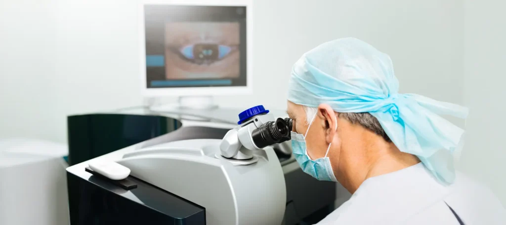
Let’s talk about something you might not hear every day: the chemical fingerprint of your eyes. More specifically, how the study of tiny molecules—known as metabolites—in and around your eye might help detect cataracts long before your vision gets cloudy. It’s all part of an exciting new field called lens metabolomics, and yes, it could completely change how we approach cataract diagnosis and prevention.
Cataracts are a leading cause of blindness globally. But what if we didn’t have to wait until the cloudiness kicks in to know something’s wrong? What if there were a subtle whisper of biochemical change—right in your tear fluid or aqueous humour—that could alert us to early lens deterioration? That’s what metabolomics is aiming to uncover.
What is Metabolomics—and Why Should You Care?
If you’re new to the term, metabolomics is the comprehensive study of small molecules (metabolites) in biological systems. Think of it like a snapshot of the body’s chemistry at a given time. Unlike genes or proteins, metabolites reflect real-time changes—how your body is functioning right now, not just what it’s capable of.
For eye health, metabolomics gives us a powerful lens (pun intended) into early disease processes. It allows researchers and clinicians to detect biochemical imbalances or oxidative stress before physical symptoms—like blurry vision or glare—ever set in. In the context of cataracts, that means spotting trouble while the lens is still transparent.
The Human Lens: A Silent Battleground
Your eye’s natural lens sits just behind the iris and helps focus light onto the retina. It’s made mostly of water and proteins arranged in a precise structure to keep it clear. But over time, exposure to UV light, oxidative stress, metabolic diseases like diabetes, and even certain medications can trigger a cascade of molecular changes.
These changes lead to the clumping of proteins, disruption of the lens architecture, and eventual clouding—that’s your cataract forming. The frustrating part? These changes begin long before you notice anything’s off. And that’s where metabolomics could step in.
Current Methods: Why They Fall Short
At the moment, most cataracts are diagnosed during a routine eye exam. Your optometrist might spot the early signs using slit-lamp imaging, but that usually happens after some structural changes are already underway.
What we don’t currently have is a simple biochemical test—a bit like a blood test—that can say, “Hey, your lens is starting to go off track, even if you’re not noticing it yet.” That’s the gap lens metabolomics is hoping to fill.
Tear Fluid and Aqueous Humour: Nature’s Diagnostic Fluids
Now, you might be wondering—how can we access what’s going on in the lens without poking around in the eye? That’s where tear fluid and aqueous humour come in.
Tear Fluid: It’s easily accessible, non-invasive to collect, and surprisingly rich in biological information. Your tears don’t just express emotion—they carry proteins, enzymes, and small molecules that reflect your eye’s internal state. That includes changes that may originate from lens metabolism.
Aqueous Humour: This is the clear fluid that fills the front part of the eye, between the lens and the cornea. While collecting it is more invasive, it offers an even closer reflection of the environment surrounding the lens. Metabolic changes that affect lens health are often mirrored here.
What We’re Finding: Early Biomarkers and Metabolic Signatures

Recent studies using mass spectrometry and nuclear magnetic resonance (NMR) spectroscopy have started to uncover specific metabolites associated with early lens ageing and cataract formation. Here are a few that are grabbing attention:
- • Glutathione and its precursors
Glutathione is one of the most vital antioxidants in the human eye, particularly in the lens where it acts as a key defender against oxidative stress. It exists in both reduced (GSH) and oxidised (GSSG) forms, helping to neutralise free radicals and maintain the transparency of the lens proteins. When glutathione levels decline, it’s a strong indicator that the lens is struggling to combat oxidative insults—be they from UV radiation, ageing, or metabolic imbalances. Studies have shown that the earliest signs of cataractogenesis often coincide with a drop in GSH levels and an altered GSH/GSSG ratio.
The body doesn’t just rely on glutathione itself but also on its precursors—like cysteine, glutamate, and glycine—to maintain healthy levels. Any disruption in the synthesis or recycling of glutathione may be reflected in tear or aqueous humour samples. This makes it a promising biomarker candidate. Detecting a decline in glutathione or its precursors in these fluids could provide an early biochemical red flag, long before structural changes in the lens occur. It’s this kind of real-time metabolic monitoring that could make proactive lens care a reality. - • Lactic acid and pyruvate ratios
The lens depends primarily on anaerobic glycolysis for its energy production, meaning it relies heavily on the breakdown of glucose to lactic acid without oxygen. During this process, the conversion between lactic acid and pyruvate is tightly regulated. An imbalance in the lactic acid-to-pyruvate ratio can indicate that the lens is undergoing metabolic stress or experiencing inefficient energy utilisation. This disruption has been observed in early cataract development, especially in diabetic patients or those exposed to chronic oxidative stress.
By examining tear fluid or aqueous humour, researchers can detect subtle shifts in this ratio that may signal early metabolic dysfunction in the lens. For example, an excess of lactic acid may suggest hypoxic stress or mitochondrial impairment—both of which can precede protein misfolding and lens opacification. These changes might not be picked up during a standard eye exam, but they can provide crucial early insights when analysed through metabolomics. This ratio, therefore, offers a valuable snapshot of the lens’s energy status and vulnerability. - • Tryptophan metabolites (kynurenines)
Tryptophan, an essential amino acid, is metabolised through several pathways—one of the most relevant to eye health being the kynurenine pathway. Kynurenines are produced in response to UV exposure and oxidative stress and can accumulate in the lens over time. In healthy eyes, they serve a protective function, absorbing UV light. But when this balance is tipped, excessive or altered forms of kynurenines can contribute to the cross-linking of lens proteins, promoting cloudiness and early cataract formation.
Elevated levels of these metabolites have been detected in the aqueous humour of patients showing early signs of lens opacity. Their presence suggests not only UV-induced damage but also ongoing protein oxidation and inflammation within the lens microenvironment. This makes kynurenines valuable as early diagnostic markers, especially for those with high sun exposure or limited antioxidant defences. Tracking these metabolites could also help tailor preventative strategies, such as targeted supplementation or lifestyle changes, aimed at preserving lens clarity. - • Ascorbate levels (Vitamin C)
Ascorbate, more commonly known as Vitamin C, is another crucial antioxidant in the eye, particularly in the aqueous humour where it protects both the lens and cornea from oxidative injury. It works by scavenging harmful free radicals generated by UV light and metabolic activity. In the lens, Vitamin C not only shields against oxidative stress but also helps regenerate other antioxidants like Vitamin E and glutathione. A deficiency in ascorbate concentration can significantly reduce the lens’s resilience to oxidative insults, accelerating the process of cataract formation.
Recent metabolomic studies have identified altered ascorbate levels in individuals at risk of early cataract development. Low ascorbate concentrations can suggest a reduced capacity for antioxidant defence, especially in the presence of diabetes, ageing, or high environmental stress. This biomarker is particularly promising because Vitamin C levels can potentially be boosted through diet or supplementation. Detecting a drop early gives clinicians a potential window to intervene and help maintain lens health before structural changes become irreversible. Researchers are now looking at these and other molecules as possible biomarkers for an early warning system.
How Could This Translate to the Clinic?
Imagine going for a simple eye test in the future—not just to measure your prescription, but to submit a small tear sample. Within minutes, it could be analysed for key metabolites that tell your optometrist whether your lens is starting to show signs of oxidative stress or protein misfolding.

From there, you might get specific lifestyle recommendations, antioxidant supplements, or earlier follow-ups—all before the first sign of vision loss. It’s proactive, personal, and potentially transformative.
The Tech Behind the Breakthrough
To do this kind of analysis, researchers use sophisticated tools like:
- • Mass Spectrometry (MS):
Mass spectrometry is a powerhouse in metabolomics, allowing scientists to detect and quantify hundreds of metabolites from incredibly small volumes of tear fluid or aqueous humour. It works by ionising chemical compounds and measuring the mass-to-charge ratio of the resulting ions, creating a precise chemical fingerprint. This enables researchers to identify which metabolites are present and in what quantities—even in samples just a few microlitres in size. Its sensitivity makes it invaluable for detecting early biochemical changes in the eye that may signal cataract development before any physical signs are visible. - • Liquid Chromatography (LC):
Liquid chromatography plays a critical supporting role by separating individual compounds in a biological sample before they are analysed by mass spectrometry. This is important because biological fluids like tears or aqueous humour contain complex mixtures of molecules that can interfere with one another if not properly isolated. LC helps ensure that each metabolite enters the mass spectrometer on its own, boosting both accuracy and resolution. This separation step is crucial for identifying subtle shifts in metabolites that could serve as early indicators of lens stress or dysfunction. - • NMR Spectroscopy:
Nuclear Magnetic Resonance (NMR) spectroscopy offers a unique advantage in metabolomics by providing detailed structural information about each metabolite in a sample. While slightly less sensitive than mass spectrometry, NMR doesn’t require extensive sample preparation and can analyse mixtures without separating them first. This makes it especially useful for identifying previously unknown compounds or confirming the identity of known biomarkers. In the context of cataract research, NMR can help pinpoint which molecular changes in the lens or surrounding fluids are most closely associated with the onset of opacification. - • Bioinformatics:
Bioinformatics is the digital engine that powers modern metabolomics, turning raw data into meaningful insights. By using advanced algorithms, statistical models, and machine learning, researchers can sift through the vast volumes of data generated by MS, LC, and NMR to detect patterns and correlations. These tools help identify which combinations of metabolites best predict early cataract formation, and which changes are most significant over time. As datasets grow, bioinformatics also enables personalised risk profiling, opening the door to highly tailored interventions based on an individual’s unique metabolic signature.
These technologies have come a long way in the past decade, enabling high-resolution metabolic fingerprinting with just microlitres of fluid.
Diabetes and Cataracts: A Perfect Case for Metabolomics
If you have diabetes, you’re at a higher risk of developing cataracts earlier and faster than non-diabetics. Why? High blood sugar affects lens metabolism and accelerates protein glycation and oxidative stress.

Tear metabolomics is already showing promise in identifying early diabetic lens changes—even before cataracts form. That means targeted interventions for diabetic patients could come sooner, potentially delaying or even preventing surgery.
Challenges Still to Overcome
Of course, we’re not quite at the point where you can walk into a high street optician and get your tears scanned. Here’s what still needs to happen:
- Standardisation of Methods: Tear and aqueous metabolite levels vary based on collection technique, time of day, and individual hydration. Researchers are working to establish consistent protocols.
- Larger Sample Sizes: Most current studies are small and experimental. We need large-scale population studies to validate biomarkers.
- Integration into Practice: Eye clinics will need affordable, rapid, and user-friendly diagnostic tools that can interpret metabolomics data in real time.
It’s not a question of if—but when.
The Role of AI and Big Data
To make sense of the thousands of metabolites in each tear sample, AI is playing a central role. Machine learning algorithms can spot patterns, classify disease stages, and even predict future risk with surprising accuracy.
This opens the door for fully integrated diagnostic platforms—where tear samples are analysed instantly, and risk scores are generated in seconds. Think of it as a biochemical early warning dashboard for your eyes.
Could We One Day Prevent Cataracts?
This is perhaps the most exciting implication of all. If metabolomics can flag the earliest changes in lens chemistry, then we’re not just talking about early detection—we’re talking about prevention.
Future treatments could include:
- Topical drops targeting specific metabolic imbalances
- Nutraceuticals designed to replenish depleted metabolites
- Lifestyle interventions tailored to your unique metabolic profile
In other words, cataracts might one day become something you manage, not something you simply wait for and then treat surgically.
What This Means for You
If you’re someone with a family history of cataracts, diabetes, or just getting older (aren’t we all?), then metabolomics is a field to watch. It’s giving us a whole new set of tools to understand eye health on a molecular level.
In the coming years, routine check-ups may include not only vision charts and pressure tests but biochemical profiling from a tear sample. The more we learn about your eye’s metabolic environment, the better we can tailor your care.

Final Thoughts: The Lens as a Window to Your Body
Your lens may be tiny, but it’s a highly sensitive barometer of systemic and local health. The same processes that affect your eyes—oxidation, glycation, inflammation—are also at play throughout your body.
By using metabolomics to track lens health, we’re also learning more about ageing, chronic disease, and how to personalise healthcare more broadly. The eye, once again, proves to be a window—not just to the soul, but to your biology.
So next time your eyes water a little, think about the treasure trove of information those drops may hold. In the world of early cataract detection, your tears might just be the clearest sign of all.
References
- Young, R.W., 1991. Pathophysiology of age-related cataract. Survey of Ophthalmology, 35(2), pp.119–134.
- Truscott, R.J.W., 2005. Age-related nuclear cataract—oxidation is the key. Experimental Eye Research, 80(5), pp.709–725.
- NHS, 2023. Cataracts – Causes. NHS.uk.
- Gibson, D.S. and Rooney, M.E., 2007. The human tear proteome as a source of biomarkers for ocular and systemic disease. Proteomics – Clinical Applications, 1(10), pp.876–885.
- Sereika, M., Redzic, Z. and Bukowy-Bieryllo, Z., 2021. Metabolomics and proteomics of the human aqueous humour and lens: Emerging insights into cataractogenesis. International Journal of Molecular Sciences, 22(3), pp.1–19.
Available at: https://www.mdpi.com/1422-0067/22/3/1280

