If you’ve ever had an injury to your eye—whether it was a knock during sport, an accident at work, or even something as small as a scratch from a tree branch—you might be surprised to learn that it could have long-term consequences. One of the most concerning complications of eye trauma is the increased risk of developing cataracts. In this article, we’ll walk through exactly how trauma can lead to cataracts, what signs to watch for, and what treatment options are available.
What Is a Cataract?
Before diving into trauma-related cataracts, let’s quickly recap what a cataract actually is. A cataract is the clouding of the eye’s natural lens, which sits just behind the iris. Normally, this lens is crystal clear, allowing light to pass through easily and focus on the retina. When a cataract forms, that clarity is lost, leading to blurry vision, glare, and difficulty with contrast and colour perception.
Cataracts are often associated with ageing, but trauma is a lesser-known yet significant cause. Traumatic cataracts can develop suddenly or gradually, depending on the type and severity of the injury.
How Eye Trauma Leads to Cataract Formation
When your eye experiences physical trauma, several changes can occur within the eye’s internal structures. These changes can disrupt the lens fibres and the delicate capsule that surrounds the lens. If the capsule is damaged, the proteins inside the lens can clump together, causing cloudiness.
There are two primary types of trauma that can result in cataracts:
1. Blunt Trauma
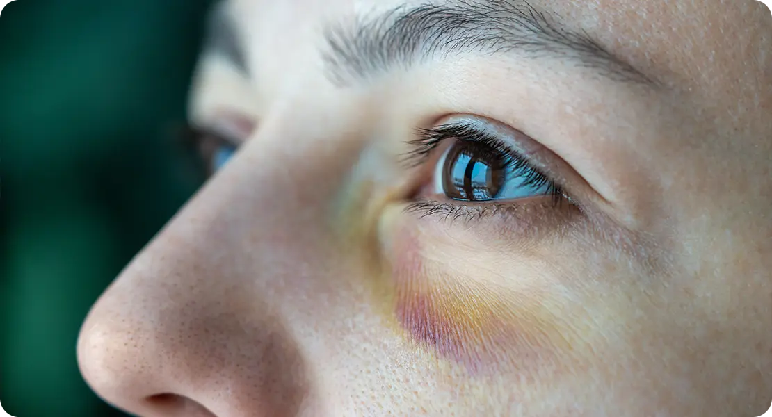
Blunt trauma refers to a forceful impact to the eye that doesn’t penetrate the surface but causes internal disruption. This can happen from a punch, a ball hitting the eye, or even something like an airbag deploying in a car accident. The energy from the blow travels through the eye, damaging structures like the lens, which is responsible for focusing light. Despite the outer appearance of the eye often remaining intact, internal damage may be significant and not immediately obvious.
A typical result of blunt trauma is a rosette-shaped cataract, which forms due to the shockwave damaging the lens fibres. These opacities often begin subtly but can expand and distort vision over time. Their unique pattern makes them recognisable during an eye examination, helping doctors link the cataract directly to a traumatic event. The visual impact of these cataracts depends on their size and location within the lens, with central opacities causing more disruption.
Injuries like these may also affect other parts of the eye that support the lens, including the zonular fibres. If these fibres are stretched or torn, the lens can become unstable or shift out of place, leading to further visual disturbances and increasing the risk of cataract development. Additionally, trauma-induced inflammation, such as iritis or uveitis, can trigger changes in lens metabolism, promoting cloudiness and deterioration of lens clarity over time.
Blunt trauma doesn’t always lead to immediate cataract formation. In many cases, the damage progresses slowly, with symptoms becoming noticeable months or even years after the original injury. This delayed development is why ongoing monitoring is so important following eye trauma. Regular check-ups can identify early changes and ensure timely management to preserve vision and plan for treatment if needed.
2. Penetrating Injury
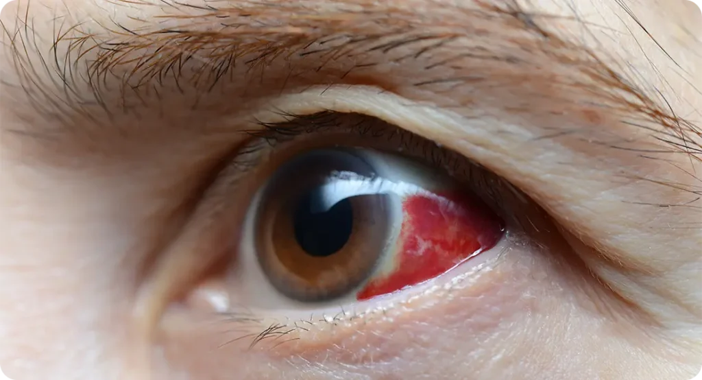
Penetrating injuries occur when a foreign object enters the eye, breaching the outer layers and often damaging internal structures, including the lens. These injuries are typically caused by sharp or fast-moving objects such as metal, glass, or wood. Because the lens is located just behind the front part of the eye, it is highly susceptible to direct trauma. Once the lens capsule is torn, its protective barrier is lost, making it prone to inflammation and rapid clouding.

Traumatic cataracts from penetrating injuries tend to form quickly. The disruption of the capsule allows fluid and inflammatory cells to mix with the lens material, which leads to swelling, breakdown of proteins, and cloudiness. The clarity of vision can decline sharply in a short period, and patients often notice a sudden onset of visual disturbance. Unlike age-related cataracts, which develop gradually, these cataracts can become visually significant within days.
There is also a high risk of infection when the eye is breached. Contaminants carried by the foreign object can introduce bacteria into the eye, potentially leading to a serious condition known as endophthalmitis. Inflammatory responses to such infections can worsen cataract formation and threaten overall eye health. Managing these situations often requires immediate antibiotic treatment alongside surgical planning.
Surgical removal of traumatic cataracts caused by penetrating injuries is often more complicated than standard procedures. The extent of structural damage may require tailored techniques and the use of alternative intraocular lens placement methods. In some cases, surgery may be delayed to allow the eye to recover from inflammation or infection. The long-term visual outcome largely depends on whether the injury has affected other parts of the eye such as the retina or optic nerve.
Recognising the Symptoms of a Traumatic Cataract
The signs of a cataract following trauma can vary. Some changes in vision might occur immediately, while others may not appear until weeks or even months after the injury. Common symptoms to look out for include:
- Blurred or cloudy vision
- Sensitivity to light or glare
- Seeing halos around lights
- Faded or yellowed colours
- Poor night vision
- Double vision in one eye
If you’ve suffered an eye injury and notice any of these symptoms, it’s essential to see an eye specialist as soon as possible.

Diagnosing a Traumatic Cataract
The diagnosis starts with a full eye examination, usually carried out by an ophthalmologist. They will assess your visual acuity and use a slit-lamp microscope to get a detailed look at the structures of your eye. The location, shape, and density of the cataract will help determine how trauma has affected the lens.
In some cases, additional imaging such as an ultrasound or OCT scan might be used—especially if the trauma has caused bleeding, swelling, or retinal detachment. It’s also important to check for other associated injuries like lens dislocation or damage to the cornea.
Treatment Options for Traumatic Cataracts
Not all traumatic cataracts need immediate surgery. If the cataract is small and not significantly affecting vision, your ophthalmologist may recommend simply monitoring it over time. However, if your vision is severely impaired, cataract surgery is usually the next step.
Cataract Surgery After Trauma
The surgical approach to removing a traumatic cataract can be more complex than routine age-related cataract surgery. Here’s why:
• The lens capsule may be damaged or torn
When trauma disrupts the integrity of the lens capsule, it significantly alters the dynamics of cataract surgery. The capsule acts as a support structure during the removal of the cataract and placement of the new lens, so any damage to it makes standard techniques riskier. A torn capsule may result in fragments of the lens dropping into the back of the eye during surgery, which complicates the procedure and can affect visual outcomes if not managed correctly.
Surgeons may need to use advanced techniques to stabilise the lens or clean up lens material from the vitreous cavity if it migrates backwards. This often involves the use of a vitrectomy—a specialised procedure to remove the gel-like substance in the back of the eye and clear any displaced lens matter. These additional steps require more time and expertise, and the need for them is typically determined intraoperatively, depending on the extent of the damage discovered.
In some cases, damage to the capsule may be so extensive that it is no longer able to support any type of artificial lens. This necessitates a more customised approach, where the surgeon must decide whether to implant a lens elsewhere in the eye or delay implantation entirely. The absence of a stable capsule also means that future surgeries may be needed to fine-tune the patient’s visual rehabilitation.
• There may be other internal injuries that need addressing during the same procedure
Eye trauma that causes cataracts often affects more than just the lens. The same incident might damage the iris, dislocate the natural lens, or cause bleeding inside the eye. These coexisting injuries complicate the surgical landscape and require a broader treatment plan than simple lens removal. It’s not uncommon for surgeons to encounter multiple issues that must be handled in a single session to give the patient the best chance of recovery.
Managing these additional injuries during cataract surgery may require the use of specialised surgical tools and techniques. For example, if the iris is torn or displaced, surgical repair may be necessary to restore the natural pupil shape and allow proper light entry. Retinal tears or detachment—often invisible without preoperative imaging—must also be identified and treated immediately to prevent vision loss. In these cases, a retina specialist may need to be involved as part of a collaborative surgical team.
The presence of bleeding, or hyphema, can obscure the surgeon’s view during the procedure, increasing the risk of complications. If blood enters the anterior chamber or vitreous cavity, it can reduce visibility and make delicate manoeuvres more challenging. This is why thorough preoperative planning, imaging, and sometimes a staged approach to surgery are required in trauma-related cataracts—each step must be coordinated to reduce risks and maximise visual outcomes.
• Sometimes a standard lens implant (intraocular lens or IOL) cannot be placed in the usual position, and alternative fixation techniques must be used
In a typical cataract operation, the artificial lens is placed securely within the capsular bag that once held the natural lens. However, in trauma cases where the capsule is ruptured or unstable, this standard placement may no longer be possible. When this happens, surgeons must use alternative methods to secure the new lens, which can include placement in the ciliary sulcus, iris fixation, or even suturing the lens to the scleral wall of the eye.

Each of these alternative techniques carries its own set of risks and benefits. Sulcus placement requires enough remaining support tissue around the lens, and improper positioning can lead to lens tilt or decentration, affecting vision quality. Iris-claw lenses, which clip onto the front or back of the iris, are a good option in select cases but may increase the risk of inflammation or damage to the iris tissue over time. Scleral-fixated lenses, which are sutured into place, provide strong long-term positioning but require meticulous surgical skill to avoid complications such as suture erosion or lens dislocation.
The decision on which method to use is influenced by the patient’s eye anatomy, the extent of trauma, and the surgeon’s experience with each technique. Postoperative care is also more complex, with increased monitoring for signs of intraocular pressure changes, lens instability, or inflammation. While these approaches are more involved than routine lens implantation, they are often highly successful when carefully executed, allowing patients to regain stable and functional vision.
Recovery and Outlook
Recovery from traumatic cataract surgery will vary depending on the severity of the original injury and any complications. You’ll likely need eye drops to reduce inflammation and prevent infection, and regular follow-up appointments to monitor healing. In many cases, vision improves significantly after the cataract is removed and the eye has had time to recover.
However, it’s important to note that the final visual outcome also depends on whether other parts of the eye—like the retina or optic nerve—were affected during the trauma.
Key Takeaways
- Trauma can damage the eye’s lens, leading to cataract formation.
- Both blunt and penetrating injuries can result in traumatic cataracts.
- Symptoms may appear immediately or develop gradually after an injury.
- Prompt diagnosis and monitoring are essential to manage long-term risks.
- Surgery is effective in most cases, but the complexity depends on the nature of the trauma.
Final Thoughts
It’s easy to dismiss an eye injury as a one-off incident, especially if your vision seems fine in the days that follow. But trauma can quietly trigger long-term changes inside the eye—cataracts being one of the most common consequences. If you’ve ever had a significant eye injury, it’s worth having regular eye checks, even years later. Early detection and intervention can make a huge difference in preserving your sight.
Have you had an eye injury in the past and are now noticing changes in your vision? Don’t wait—speak to your optometrist or ophthalmologist and get your eyes properly checked. Your future vision may depend on it. If you’re concerned that your symptoms might be linked to cataracts caused by trauma, you’re welcome to contact us at the London Cataract Centre to arrange a consultation with one of our expert cataract surgeons.

