")

Cataracts remain one of the leading causes of visual impairment globally, and among the various types, posterior subcapsular cataracts (PSCs) present unique clinical features, risk factors, and treatment considerations. PSCs are particularly significant due to their disproportionate impact on visual acuity, especially in bright light or while reading. Unlike nuclear or cortical cataracts, PSCs develop at the back of the lens, just in front of the posterior capsule, in the path of light entering the eye. This article explores the pathophysiology, clinical presentation, risk factors, diagnosis, and management of posterior subcapsular cataracts.
Pathophysiology
The lens of the eye is composed of concentric layers of fibres, with the outer layers forming the cortex and the central zone comprising the nucleus. The posterior subcapsular area is located just anterior to the lens capsule at the back of the lens. PSCs form when abnormal migration and proliferation of epithelial cells occur in this region, resulting in the formation of granular or plaque-like opacities.
These opacities are composed of aberrant lens fibres and protein aggregates, which scatter incoming light and significantly reduce visual clarity. Because they are located directly in the visual axis, even small PSCs can lead to considerable visual symptoms, in contrast to other types of cataracts where larger opacities are needed to cause noticeable vision problems.
Risk Factors
Prolonged corticosteroid use
Corticosteroids, whether administered systemically or topically, have long been associated with the development of posterior subcapsular cataracts. Their role in altering lens cell physiology is believed to contribute significantly to this process. Corticosteroids can disrupt the balance of electrolytes and proteins within the lens fibres, leading to the aggregation of proteins and clouding at the posterior aspect. These changes may occur insidiously, even with relatively low doses taken over an extended period, making long-term corticosteroid therapy a concern in chronic conditions such as asthma, autoimmune diseases, and post-transplant immunosuppression.
Moreover, topical steroid use—particularly in the form of eye drops for conditions like uveitis or allergic conjunctivitis—can also lead to PSC formation, especially when used without close monitoring. The lens’s posterior region is particularly susceptible to metabolic disruptions caused by steroids due to its low regenerative capacity and high metabolic demand. This makes it critical for healthcare professionals to weigh the risks and benefits of prolonged corticosteroid therapy and explore alternatives or adjunctive therapies when possible to mitigate ocular complications.
Diabetes mellitus
Diabetes mellitus is another major systemic contributor to the development of posterior subcapsular cataracts. The persistent hyperglycaemic state associated with poorly controlled diabetes can lead to the accumulation of sorbitol within the lens, a by-product of glucose metabolism via the polyol pathway. This osmotic imbalance causes lens fibre swelling, oxidative stress, and eventual damage, particularly affecting the posterior subcapsular area due to its high vulnerability to such metabolic insults.

In addition, individuals with diabetes often exhibit earlier and more aggressive cataract formation than the general population, with PSCs being a frequent manifestation. The interplay between diabetic microvascular changes and lens metabolism creates an environment conducive to cataractogenesis. Regular ophthalmic evaluations are crucial for diabetic patients, not only to detect early cataract changes but also to manage co-existing retinal complications such as diabetic retinopathy, which may influence the timing and outcomes of cataract surgery.
Radiation exposure
Exposure to ionising radiation is a well-documented risk factor for PSC development, particularly in individuals undergoing radiotherapy for head and neck malignancies. Radiation affects the DNA and cellular structures of lens epithelial cells, prompting abnormal cell migration and protein deposition in the posterior subcapsular region. The damage is typically cumulative and may not manifest immediately, sometimes taking months or even years after exposure for symptoms to appear.
Importantly, the risk is not confined to therapeutic radiation alone. Occupational exposure, such as in healthcare workers dealing with radiological equipment or airline crew exposed to cosmic rays, can also contribute over time. Protective measures, including shielding and adherence to radiation safety protocols, are essential in these settings. Even environmental factors, like prolonged UV exposure without eye protection, may exert a contributory effect due to the oxidative stress imposed on the lens fibres.
Younger age
Unlike other cataract types that are generally associated with ageing, posterior subcapsular cataracts are often seen in younger individuals. This is particularly evident in patients who have chronic illnesses requiring long-term steroid use or those with inflammatory or metabolic disorders from a young age. The early onset of PSCs can significantly affect quality of life, particularly when the demands of reading, driving, and screen usage are high.
The psychosocial impact of PSCs in younger populations should not be underestimated. Many of these patients may require cataract surgery at an earlier age, raising concerns about intraocular lens longevity, the potential for posterior capsule opacification, and the need for future interventions. Tailored follow-up and a multidisciplinary approach are essential to manage these patients, especially in cases where cataract formation is linked to a chronic systemic condition.
Ocular inflammation
Chronic or recurrent ocular inflammation, such as that seen in uveitis, can accelerate the development of PSCs. Inflammatory mediators like cytokines and prostaglandins can disrupt the homeostasis of the lens microenvironment, leading to cellular damage and the accumulation of opacities. Moreover, the treatment of uveitis often includes corticosteroids, which compound the risk, making the lens doubly susceptible to cataract formation.
Management of these patients can be particularly challenging due to the ongoing risk of inflammation. Cataract surgery in the presence of uveitis must be timed carefully, ideally during a quiescent phase of disease to minimise the risk of postoperative complications such as cystoid macular oedema or exacerbation of inflammation. Coordinated care between ophthalmologists and rheumatologists or immunologists is often required to optimise both ocular and systemic outcomes.
Genetic predisposition
A growing body of evidence suggests that genetic factors may play a role in the development of PSCs, although the exact mechanisms remain under investigation. Mutations or polymorphisms in genes regulating lens transparency, oxidative defence systems, or epithelial cell behaviour could predispose individuals to early or accelerated cataract formation. Familial clustering of PSCs has been reported in some studies, hinting at hereditary components that are yet to be fully elucidated.
Furthermore, PSCs are sometimes associated with specific genetic syndromes such as Down syndrome or myotonic dystrophy, where cataract formation occurs at a much younger age. Identifying genetic predispositions can help guide screening protocols, particularly for individuals with a strong family history of early cataract surgery. Advances in genetic testing may eventually allow for more personalised monitoring and early intervention strategies.
Clinical Features
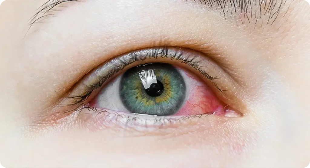
Glare and light sensitivity
One of the hallmark symptoms of posterior subcapsular cataracts is increased glare sensitivity, which patients often describe as an overwhelming brightness or difficulty seeing in sunlight or under strong artificial lighting. This is due to the central and posterior location of the opacity, which interferes with the passage of light directly along the visual axis. Light entering the eye becomes scattered as it hits the irregular surface of the cataract, leading to a sensation of haze and discomfort, especially when the pupil constricts in bright environments.
This symptom can be particularly disabling for those who drive at night, as the glare from oncoming headlights may create halos or starbursts, significantly reducing contrast sensitivity. In many cases, patients might find that their vision improves in dim lighting or when their pupils are dilated, reducing the light’s direct contact with the affected area. However, this adaptation is not sufficient for daily functioning, prompting many to seek medical attention even before a major decline in visual acuity is noted.
Difficulty reading or performing near tasks
Patients with PSCs frequently report difficulty with tasks that require focused vision, such as reading, writing, or using digital screens. This issue arises because PSCs tend to impair near vision more acutely than distance vision. The dense central opacity disrupts the convergence of light rays necessary for forming a clear image on the retina, leading to blurred or shadowed vision at close range. Even with glasses or magnifiers, clarity may remain elusive.

This challenge often becomes more frustrating for patients who previously had good near vision, especially in those who are still active in the workforce or pursuing education. The inconsistency of vision—clear at times and blurry at others—can cause fatigue, reduced concentration, and diminished productivity. These issues may lead individuals to believe their glasses are no longer working, when in fact the root cause lies in the cataract itself.
Rapid decline in vision compared to other cataract types
Unlike nuclear or cortical cataracts, which often progress slowly over several years, PSCs are notorious for their rapid deterioration of vision. This quick onset can catch patients and clinicians off guard, especially if regular eye exams are not in place. The opacity’s central location means that even a small increase in density can lead to a noticeable drop in visual function, including reading ability, facial recognition, and visual clarity at all distances.
This rapid decline often necessitates earlier surgical intervention, as the loss of functional vision interferes significantly with daily life. Patients may struggle with tasks they managed with ease only a few months prior. Early referral and prompt treatment are essential, especially in individuals who are still driving or working. Recognising the signs early allows for timely surgery, often restoring vision to a remarkable degree.
Diagnosis
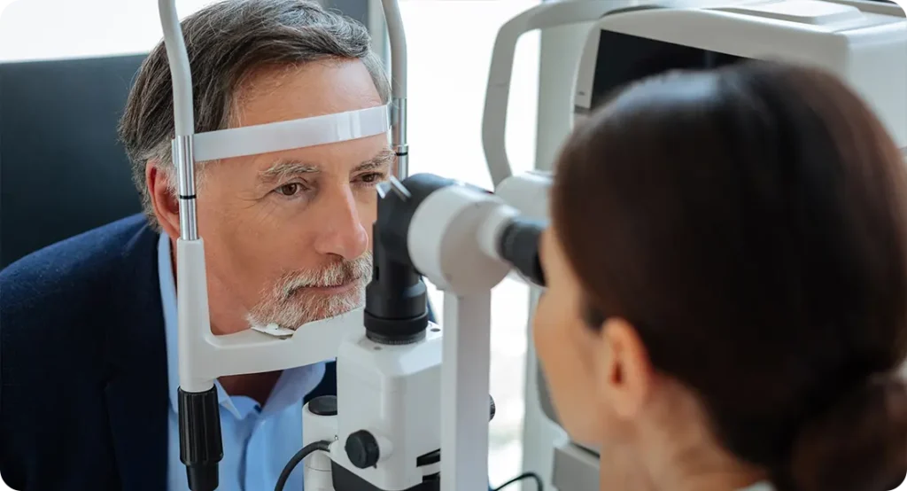
Diagnosing posterior subcapsular cataracts is typically a straightforward process, relying primarily on slit-lamp biomicroscopy—a standard tool in ophthalmic examinations. Under high magnification and appropriate illumination, PSCs can be identified as distinct, granular or plaque-like opacities situated at the back of the lens. These changes are usually localised centrally, directly along the visual axis, which helps explain their significant impact on visual function. The granular appearance may vary from fine dots to more confluent patches, depending on the severity and progression of the cataract. Careful focusing at the level of the posterior lens surface is essential to distinguish PSCs from other lens opacities, such as cortical spokes or nuclear sclerosis.
Retroillumination, often performed during the slit-lamp exam, enhances visualisation of these opacities by projecting light through the pupil and reflecting it off the retina. This technique highlights the PSC as a dark or greyish shadow silhouetted against the red reflex, making even subtle changes more apparent. Retroillumination is particularly useful for documenting progression over time and for preoperative assessments prior to cataract surgery. In some cases, patients may not fully appreciate the degree of lens opacity until it’s demonstrated with this imaging technique, which can be a valuable tool in discussing treatment options.
Additional diagnostic tools include:
- Visual acuity testing under different lighting conditions.
- Pinhole testing, which often fails to significantly improve vision in PSC due to the central obstruction.
- Ocular coherence tomography (OCT) is not required for diagnosis but may be used to assess other co-existing posterior segment conditions.
Treatment
The mainstay of treatment for posterior subcapsular cataracts is surgical intervention once the symptoms begin to interfere with daily tasks such as reading, driving, or working. Due to the location of the opacity at the back of the lens, patients with PSCs often notice visual disturbances sooner and more severely than those with other cataract types. As a result, the decision to proceed with surgery is often made earlier in the disease course. Unlike nuclear cataracts that may progress gradually over years, PSCs tend to deteriorate visual function more quickly, prompting more urgent management.
While non-surgical options such as optimised lighting, magnifying aids, or updated spectacle prescriptions may offer temporary relief, they are unlikely to provide long-term benefit. This is because the opacity lies directly in the visual axis, so even minor changes can significantly affect central vision. Therefore, conservative management is usually limited to patients with mild symptoms or those not yet ready for surgery due to other health factors.
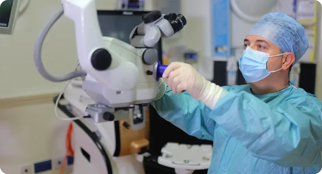
Phacoemulsification remains the gold standard, involving the removal of the opacified lens and replacement with an artificial intraocular lens (IOL).
Phacoemulsification is a minimally invasive surgical technique that uses ultrasonic energy to break the cloudy lens into tiny fragments, which are then aspirated from the eye. A foldable intraocular lens is implanted in the empty capsular bag, effectively restoring clarity by replacing the eye’s natural focusing element. This method allows for a small incision, typically self-sealing, which minimises healing time and reduces the risk of complications.
The advantages of phacoemulsification are particularly evident in PSCs due to their tendency to affect patients at a younger age or in the presence of co-morbid conditions. The precision and control offered by modern equipment help ensure accurate lens positioning and excellent visual outcomes. Patients often experience significant improvements in vision within days of surgery, although full recovery may take a few weeks. As with any surgery, the success of the procedure depends on careful technique, appropriate patient selection, and postoperative care.
Preoperative planning should take into account any co-morbid conditions, such as diabetes or uveitis, which may complicate recovery.
Thorough preoperative assessment is essential to optimise surgical outcomes, particularly in patients with systemic or ocular co-morbidities. For example, poorly controlled diabetes may increase the risk of postoperative inflammation, delayed healing, or macular oedema, all of which can impact visual recovery. Similarly, in patients with a history of uveitis, inflammation must be well-controlled prior to surgery to reduce the risk of flare-ups that could compromise surgical results.
In addition to medical history, preoperative evaluations typically include biometry to determine the correct power of the IOL, ocular surface assessment, and detailed fundus examination. Where the posterior segment is not clearly visible due to the density of the cataract, ocular imaging such as B-scan ultrasonography may be needed to rule out retinal or optic nerve disease. Informed consent should address the potential for complications and the need for ongoing management of underlying conditions, even after successful cataract removal.
Postoperative visual outcomes are typically excellent, provided there are no underlying retinal or optic nerve pathologies.
Following cataract surgery for PSCs, most patients enjoy a significant and rapid improvement in visual clarity, often surpassing expectations due to the dramatic contrast between pre- and postoperative vision. The central location of the opacity means that its removal offers immediate relief from issues like glare, blurriness, and reading difficulty. The restoration of contrast sensitivity and visual sharpness can dramatically improve quality of life, particularly in those whose daily functioning had been compromised.
However, the overall success of the procedure hinges on the health of the posterior segment. If the retina or optic nerve is damaged due to pre-existing conditions—such as diabetic retinopathy, glaucoma, or macular degeneration—the visual outcome may be limited despite a technically flawless surgery. For this reason, realistic expectations must be set through careful evaluation and counselling. Follow-up care is also vital to monitor healing, manage intraocular pressure, and detect early signs of complications like posterior capsule opacification, which may develop months or years after the operation.
Medical treatment, such as adjusting corticosteroid use or managing diabetes more tightly, may help slow progression but cannot reverse existing opacities. Although these measures won’t clear an existing PSC, they play a critical role in delaying further lens damage and preserving remaining vision until surgery becomes necessary. For patients not yet ready for surgery, optimising systemic health can buy valuable time while also improving surgical safety when the time comes.
In addition, reducing or tapering steroid therapy—where medically appropriate—can prevent the worsening of lens opacities. Any adjustments should be made in coordination with the prescribing specialist to avoid exacerbating the underlying condition. Similarly, encouraging tight glycaemic control in diabetic patients may slow the oxidative damage to the lens, potentially reducing the speed at which the cataract progresses. These supportive measures are not alternatives to surgery but rather part of a holistic approach to managing the patient’s visual health.
Prognosis
Following cataract surgery for posterior subcapsular cataracts, the majority of patients report a marked improvement in their vision. Colours appear more vivid, contrast is enhanced, and the bothersome glare and blurriness that once interfered with daily tasks typically vanish. This transformation often leads to a noticeable boost in overall quality of life, allowing individuals to return to reading, driving, or working with greater confidence and ease. Many patients are surprised at how much clearer their world becomes once the cloudy lens is removed and replaced with a high-quality intraocular lens (IOL). Although the healing process can vary from person to person, most regain excellent visual function within weeks of the procedure.
Despite these positive outcomes, one potential postoperative complication is posterior capsule opacification (PCO), often referred to as a “secondary cataract.” This condition arises when residual lens epithelial cells proliferate on the capsule that holds the IOL, causing it to become cloudy over time. PCO is particularly common after surgery for PSCs due to the posterior location of the original cataract and the closer involvement of the capsule during removal. Fortunately, treatment is straightforward and effective. A quick, painless outpatient procedure known as YAG laser capsulotomy uses a focused laser to create a small opening in the cloudy capsule, restoring the clear visual pathway. Most patients notice an immediate improvement, and the effects are typically long-lasting with minimal risk of further complications.
Conclusion
Posterior subcapsular cataracts represent a distinct subtype of lens opacities that disproportionately affect vision due to their anatomical location. Early recognition is crucial, especially in at-risk individuals such as steroid users and patients with diabetes. With modern surgical techniques, the prognosis following intervention is highly favourable. As awareness increases and diagnostic tools improve, timely treatment of PSCs can lead to excellent visual outcomes and enhanced quality of life for patients.
If you are concerned about cataracts or would like to discuss your symptoms, you can contact us at the London Cataract Centre to arrange a consultation with one of our specialist cataract surgeons.